 Movie
Movie Controller

[English] 日本語
 Yorodumi
- PDB-2q3i: Crystal structure of the D10-P3/IQN17 complex: a D-peptide inhibi... -
+ Open data

- Basic information
| Entry | Database: PDB / ID: 2q3i | ||||||
|---|---|---|---|---|---|---|---|
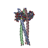
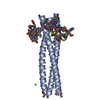
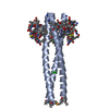
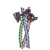
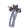
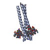
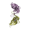
| Title | Crystal structure of the D10-P3/IQN17 complex: a D-peptide inhibitor of HIV-1 entry bound to the GP41 coiled-coil pocket | ||||||
 Components Components |
| ||||||
 Keywords Keywords | VIRAL PROTEIN/INHIBITOR / envelope glycoprotein / VIRAL PROTEIN-INHIBITOR complex | ||||||
| Function / homology |  Function and homology information Function and homology informationsymbiont-mediated perturbation of host defense response / positive regulation of plasma membrane raft polarization / positive regulation of receptor clustering / host cell endosome membrane / clathrin-dependent endocytosis of virus by host cell / viral protein processing / fusion of virus membrane with host plasma membrane / fusion of virus membrane with host endosome membrane / viral envelope / virion attachment to host cell ...symbiont-mediated perturbation of host defense response / positive regulation of plasma membrane raft polarization / positive regulation of receptor clustering / host cell endosome membrane / clathrin-dependent endocytosis of virus by host cell / viral protein processing / fusion of virus membrane with host plasma membrane / fusion of virus membrane with host endosome membrane / viral envelope / virion attachment to host cell / host cell plasma membrane / virion membrane / structural molecule activity / membrane Similarity search - Function | ||||||
| Method |  X-RAY DIFFRACTION / SYNCHROTRON / MOLECULAR REPLACEMENT / Resolution: 1.5 Å X-RAY DIFFRACTION / SYNCHROTRON / MOLECULAR REPLACEMENT / Resolution: 1.5 Å | ||||||
 Authors Authors | Malashkevich, V.N. / Eckert, D.M. / Hong, L.H. / Carr, P.A. / Kim, P.S. | ||||||
 Citation Citation | Journal: Cell(Cambridge,Mass.) / Year: 1999 Title: Inhibiting HIV Entry: Discovery of D-Peptide Inhibitors that Target the Gp41 Coiled-Coil Pocket Authors: Eckert, D.M. / Malashkevich, V.N. / Hong, L.H. / Carr, P.A. / Kim, P.S. #1: Journal: J.Mol.Biol. / Year: 1998Title: Crystal Structure of GCN4-Piqi, a Trimeric Coiled-Coil with Buried Polar Residues. Authors: Eckert, D.M. / Malashkevich, V.N. / Kim, P.S. #2: Journal: Cell(Cambridge,Mass.) / Year: 1997Title: Core structure of gp41 from the HIV envelope glycoprotein Authors: Chan, D.C. / Fass, D. / Berger, J.M. / Kim, P.S. | ||||||
| History |
|
- Structure visualization
| Structure viewer | Molecule: MolmilJmol/JSmol |
|---|
- Downloads & links
-Download
| PDBx/mmCIF format | 2q3i.cif.gz | 30.4 KB | Display | PDBx/mmCIF format |
|---|---|---|---|---|
| PDB format | pdb2q3i.ent.gz | 19.4 KB | Display | PDB format |
| PDBx/mmJSON format | 2q3i.json.gz | Tree view | PDBx/mmJSON format | |
| Others |  Other downloads Other downloads |
-Validation report
| Arichive directory | https://data.pdbj.org/pub/pdb/validation_reports/q3/2q3iftp://data.pdbj.org/pub/pdb/validation_reports/q3/2q3i | HTTPS FTP |
|---|
-Related structure data
| Related structure data |  1czqSC  2q5uC  2q7cC S: Starting model for refinement C: citing same article ( |
|---|---|
| Similar structure data |


-Links
 PDBj
PDBj


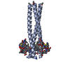
- Assembly
| Deposited unit | 
| ||||||||||||||||||
|---|---|---|---|---|---|---|---|---|---|---|---|---|---|---|---|---|---|---|---|
| 1 | 
| ||||||||||||||||||
| Unit cell |
| ||||||||||||||||||
| Components on special symmetry positions |
| ||||||||||||||||||
| Details | The biological assembly is trimer formed around the crystallographic 3-fold axis |
-Components
| #1: Protein/peptide | Mass: 5468.566 Da / Num. of mol.: 1 / Source method: obtained synthetically Details: Peptide synthesis. GCN4-HIV gp41 fusion, called IQN17. The sequence naturally occurs in Saccharomyces cerevisiae and human immunodeficiency virus References: UniProt: A3F986 |
|---|---|
| #2: Protein/peptide | Mass: 1766.993 Da / Num. of mol.: 1 / Source method: obtained synthetically / Details: Peptide synthesis. D-peptide found by screening. |
| #3: Chemical | ChemComp-CL /   Mass: 35.453 Da / Num. of mol.: 1 / Source method: obtained synthetically / Formula: Cl Mass: 35.453 Da / Num. of mol.: 1 / Source method: obtained synthetically / Formula: Cl |
| #4: Water | ChemComp-HOH /  Mass: 18.015 Da / Num. of mol.: 154 / Source method: isolated from a natural source / Formula: H2O Mass: 18.015 Da / Num. of mol.: 154 / Source method: isolated from a natural source / Formula: H2O |
| Has protein modification | Y |
-Experimental details
-Experiment
| Experiment | Method: X-RAY DIFFRACTION / Number of used crystals: 1 |
|---|
- Sample preparation
| Crystal | Density Matthews: 3.51 Å3/Da / Density % sol: 64.91 % |
|---|---|
| Crystal grow | Method: vapor diffusion / pH: 7.5 Details: 10% ETHANOL, 1.5 M SODIUM CHLORIDE, pH 7.5, VAPOR DIFFUSION |
-Data collection
| Diffraction | Mean temperature: 100 K |
|---|---|
| Diffraction source | Source: SYNCHROTRON / Site: NSLS  / Beamline: X4A / Wavelength: 0.979 Å / Beamline: X4A / Wavelength: 0.979 Å |
| Detector | Type: ADSC QUANTUM / Detector: CCD / Date: Mar 31, 2000 |
| Radiation | Protocol: SINGLE WAVELENGTH / Monochromatic (M) / Laue (L): M / Scattering type: x-ray |
| Radiation wavelength | Wavelength: 0.979 Å / Relative weight: 1 |
| Reflection | Resolution: 1.5→35 Å / Num. obs: 16309 / % possible obs: 97.2 % / Observed criterion σ(I): -3 / Redundancy: 6.5 % / Biso Wilson estimate: 21.6 Å2 / Rmerge(I) obs: 0.048 / Net I/σ(I): 16.8 |
| Reflection shell | Resolution: 1.5→1.55 Å / Redundancy: 6.1 % / Rmerge(I) obs: 0.235 / Mean I/σ(I) obs: 4.5 / % possible all: 86.8 |
- Processing
| Software |
| ||||||||||||||||||||||||||||||||||||
|---|---|---|---|---|---|---|---|---|---|---|---|---|---|---|---|---|---|---|---|---|---|---|---|---|---|---|---|---|---|---|---|---|---|---|---|---|---|
| Refinement | Method to determine structure: MOLECULAR REPLACEMENT Starting model: PDB entry 1CZQ Resolution: 1.5→10 Å / Rfactor Rfree error: 0.007 / Isotropic thermal model: RESTRAINED / Cross valid method: THROUGHOUT / σ(F): 0 / Stereochemistry target values: Engh & Huber
| ||||||||||||||||||||||||||||||||||||
| Solvent computation | Solvent model: FLAT MODEL / Bsol: 111.56 Å2 / ksol: 0.42 e/Å3 | ||||||||||||||||||||||||||||||||||||
| Displacement parameters | Biso mean: 28.3 Å2
| ||||||||||||||||||||||||||||||||||||
| Refine analyze |
| ||||||||||||||||||||||||||||||||||||
| Refinement step | Cycle: LAST / Resolution: 1.5→10 Å
| ||||||||||||||||||||||||||||||||||||
| Refine LS restraints |
| ||||||||||||||||||||||||||||||||||||
| LS refinement shell | Resolution: 1.5→1.59 Å / Rfactor Rfree error: 0.018 / Total num. of bins used: 6
| ||||||||||||||||||||||||||||||||||||
| Xplor file |
|
