- PDB-2q0j: Structure of Pseudomonas Quinolone Signal Response Protein PqsE -
+
Open data
ID or keywords:
Loading...
-
Basic information
Entry
Database: PDB / ID: 2q0j
Title
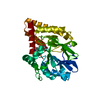
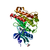
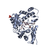
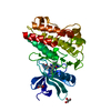
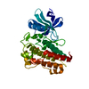
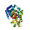
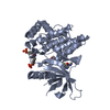
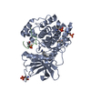
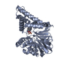
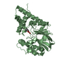
Structure of Pseudomonas Quinolone Signal Response Protein PqsE
Components
Quinolone signal response protein
Keywords
METAL BINDING PROTEIN / quorum sensing / Pseudomonas Quinolone Signal / PQS / metall-beta-lactamase / iron / phosphodiesterase
Function / homology
Function and homology information
2-aminobenzoylacetyl-CoA thioesterase / secondary metabolite biosynthetic process / hydrolase activity / metal ion binding Similarity search - Function
Type: MARMOSAIC 225 mm CCD / Detector: CCD / Date: Feb 12, 2006
Radiation
Monochromator: Si(111) / Protocol: SINGLE WAVELENGTH / Monochromatic (M) / Laue (L): M / Scattering type: x-ray
Radiation wavelength
Wavelength: 0.9786 Å / Relative weight: 1
Reflection
Resolution: 2.1→19.82 Å / Num. obs: 36642 / % possible obs: 97.8 % / Observed criterion σ(I): -3 / Biso Wilson estimate: 32.976 Å2 / Rmerge(I) obs: 0.071 / Net I/σ(I): 13.73
Reflection shell
Resolution: 2.1→2.2 Å / Rmerge(I) obs: 0.357 / Mean I/σ(I) obs: 4.6 / Num. measured obs: 14165 / Num. unique all: 4717 / % possible all: 97.5
-
Processing
Software
Name
Version
Classification
NB
XSCALE
datascaling
REFMAC
5.2.0019
refinement
PDB_EXTRACT
2
dataextraction
XDS
datareduction
PHASER
phasing
Refinement
Method to determine structure: MOLECULAR REPLACEMENT / Resolution: 2.1→19.82 Å / Cor.coef. Fo:Fc: 0.966 / Cor.coef. Fo:Fc free: 0.934 / SU B: 9.336 / SU ML: 0.113 / Cross valid method: THROUGHOUT / σ(F): 0 / ESU R: 0.201 / ESU R Free: 0.176 / Stereochemistry target values: MAXIMUM LIKELIHOOD Details: HYDROGENS HAVE BEEN ADDED IN THE RIDING POSITIONS, TLS-REFINEMENT HAS BEEN USED THROUGHOUT
Rfactor
Num. reflection
% reflection
Selection details
Rfree
0.211
1854
5.1 %
RANDOM
Rwork
0.151
-
-
-
obs
0.154
36641
97.96 %
-
Solvent computation
Ion probe radii: 0.8 Å / Shrinkage radii: 0.8 Å / VDW probe radii: 1.2 Å / Solvent model: BABINET MODEL WITH MASK
Displacement parameters
Biso mean: 25.76 Å2
Refinement step
Cycle: LAST / Resolution: 2.1→19.82 Å
Protein
Nucleic acid
Ligand
Solvent
Total
Num. atoms
4854
0
22
376
5252
Refine LS restraints
Refine-ID
Type
Dev ideal
Dev ideal target
Number
X-RAY DIFFRACTION
r_bond_refined_d
0.019
0.021
4991
X-RAY DIFFRACTION
r_bond_other_d
0
0.02
3443
X-RAY DIFFRACTION
r_angle_refined_deg
1.731
1.967
6779
X-RAY DIFFRACTION
r_angle_other_deg
4.175
3
8276
X-RAY DIFFRACTION
r_dihedral_angle_1_deg
6.366
5
611
X-RAY DIFFRACTION
r_dihedral_angle_2_deg
34.402
22.48
246
X-RAY DIFFRACTION
r_dihedral_angle_3_deg
16.044
15
821
X-RAY DIFFRACTION
r_dihedral_angle_4_deg
19.31
15
59
X-RAY DIFFRACTION
r_chiral_restr
0.11
0.2
733
X-RAY DIFFRACTION
r_gen_planes_refined
0.008
0.02
5620
X-RAY DIFFRACTION
r_gen_planes_other
0.014
0.02
1074
X-RAY DIFFRACTION
r_nbd_refined
0.236
0.2
1115
X-RAY DIFFRACTION
r_nbd_other
0.26
0.2
3651
X-RAY DIFFRACTION
r_nbtor_refined
0.18
0.2
2340
X-RAY DIFFRACTION
r_nbtor_other
0.11
0.2
2527
X-RAY DIFFRACTION
r_xyhbond_nbd_refined
0.198
0.2
337
X-RAY DIFFRACTION
r_metal_ion_refined
0.053
0.2
3
X-RAY DIFFRACTION
r_symmetry_vdw_refined
0.537
0.2
13
X-RAY DIFFRACTION
r_symmetry_vdw_other
0.349
0.2
78
X-RAY DIFFRACTION
r_symmetry_hbond_refined
0.259
0.2
14
X-RAY DIFFRACTION
r_mcbond_it
0.901
1.5
3048
X-RAY DIFFRACTION
r_mcbond_other
0
1.5
1239
X-RAY DIFFRACTION
r_mcangle_it
1.51
2
4844
X-RAY DIFFRACTION
r_scbond_it
2.798
3
1943
X-RAY DIFFRACTION
r_scangle_it
4.003
4.5
1935
LS refinement shell
Resolution: 2.1→2.154 Å / Total num. of bins used: 20
Rfactor
Num. reflection
% reflection
Rfree
0.236
140
-
Rwork
0.161
2517
-
obs
-
2657
97.47 %
Refinement TLS params.
Method: refined / Refine-ID: X-RAY DIFFRACTION
ID
L11 (°2)
L12 (°2)
L13 (°2)
L22 (°2)
L23 (°2)
L33 (°2)
S11 (Å °)
S12 (Å °)
S13 (Å °)
S21 (Å °)
S22 (Å °)
S23 (Å °)
S31 (Å °)
S32 (Å °)
S33 (Å °)
T11 (Å2)
T12 (Å2)
T13 (Å2)
T22 (Å2)
T23 (Å2)
T33 (Å2)
Origin x (Å)
Origin y (Å)
Origin z (Å)
1
0.687
-0.1526
0.0435
0.6411
0.1946
0.9432
0
-0.0393
-0.0288
-0.0929
-0.0105
0.0955
0.0109
-0.0909
0.0104
-0.056
-0.0135
0.0046
0.0012
0.0027
-0.0599
12.849
2.1993
7.0219
2
0.7439
-0.0979
-0.101
0.9154
0.0036
2.1341
-0.073
-0.101
0.0249
0.1989
0.1205
-0.0048
-0.0297
0.0175
-0.0476
-0.0526
0.0263
0.0292
0.0374
0.0063
-0.1481
24.9824
4.1263
49.0682
3
0.2596
-0.0493
0.0998
0.3975
0.1294
0.6245
0.0007
-0.0297
-0.0286
-0.012
0.0168
0.0813
0.0009
-0.032
-0.0175
-0.0672
-0.0248
0.0416
0.0039
0.0179
-0.0675
18.1906
2.2859
21.0519
Refinement TLS group
ID
Refine-ID
Refine TLS-ID
Auth asym-ID
Label asym-ID
Auth seq-ID
Label seq-ID
1
X-RAY DIFFRACTION
1
A
A
-8 - 301
12 - 321
2
X-RAY DIFFRACTION
2
B
B
1 - 301
21 - 321
3
X-RAY DIFFRACTION
3
A
I
999 - 1229
1 - 231
4
X-RAY DIFFRACTION
3
B
J
999 - 1143
1 - 145
+
About Yorodumi
-
News
-
Feb 9, 2022. New format data for meta-information of EMDB entries
New format data for meta-information of EMDB entries
Version 3 of the EMDB header file is now the official format.
The previous official version 1.9 will be removed from the archive.
In the structure databanks used in Yorodumi, some data are registered as the other names, "COVID-19 virus" and "2019-nCoV". Here are the details of the virus and the list of structure data.
Jan 31, 2019. EMDB accession codes are about to change! (news from PDBe EMDB page)
EMDB accession codes are about to change! (news from PDBe EMDB page)
The allocation of 4 digits for EMDB accession codes will soon come to an end. Whilst these codes will remain in use, new EMDB accession codes will include an additional digit and will expand incrementally as the available range of codes is exhausted. The current 4-digit format prefixed with “EMD-” (i.e. EMD-XXXX) will advance to a 5-digit format (i.e. EMD-XXXXX), and so on. It is currently estimated that the 4-digit codes will be depleted around Spring 2019, at which point the 5-digit format will come into force.
The EM Navigator/Yorodumi systems omit the EMD- prefix.
Related info.:Q: What is EMD? / ID/Accession-code notation in Yorodumi/EM Navigator
Yorodumi is a browser for structure data from EMDB, PDB, SASBDB, etc.
This page is also the successor to EM Navigator detail page, and also detail information page/front-end page for Omokage search.
The word "yorodu" (or yorozu) is an old Japanese word meaning "ten thousand". "mi" (miru) is to see.
Related info.:EMDB / PDB / SASBDB / Comparison of 3 databanks / Yorodumi Search / Aug 31, 2016. New EM Navigator & Yorodumi / Yorodumi Papers / Jmol/JSmol / Function and homology information / Changes in new EM Navigator and Yorodumi
 Movie
Movie Controller


 Open data
 Basic information
 Components
 Keywords
 Function and homology information
 Pseudomonas aeruginosa (bacteria)
 X-RAY DIFFRACTION /
  Authors
 Citation
 Structure visualization
 Downloads & links
 Other downloads
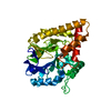

 PDBj
PDBj

 Assembly
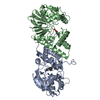

 Mass: 55.845 Da / Num. of mol.: 4 / Source method: obtained synthetically / Formula: Fe
 Mass: 122.121 Da / Num. of mol.: 2 / Source method: obtained synthetically / Formula: C7H6O2
 Mass: 18.015 Da / Num. of mol.: 376 / Source method: isolated from a natural source / Formula: H2O
 Sample preparation
 / Beamline: X10SA / Wavelength: 0.9786 Å
 Processing