 Movie
Movie Controller

[English] 日本語
 Yorodumi
- PDB-2puv: The crystal structure of isomerase domain of glucosamine-6-phosph... -
+ Open data

- Basic information
| Entry | Database: PDB / ID: 2puv | ||||||
|---|---|---|---|---|---|---|---|
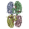
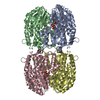
| Title | The crystal structure of isomerase domain of glucosamine-6-phosphate synthase from Candida albicans | ||||||
 Components Components | isomerase domain of glutamine-fructose-6-phosphate transaminase (isomerizing) | ||||||
 Keywords Keywords | TRANSFERASE / glucosamine-6-phosphate synthase / aldose/ketose isomerase / crystal structure / Rossmann-like fold | ||||||
| Function / homology |  Function and homology information Function and homology informationD-glucosamine biosynthetic process / hyphal growth / glutamine-fructose-6-phosphate transaminase (isomerizing) / L-glutamine:D-fructose-6-phosphate transaminase (isomerizing) activity / UDP-N-acetylglucosamine metabolic process / chitin biosynthetic process / UDP-N-acetylglucosamine biosynthetic process / carbohydrate derivative binding / fructose 6-phosphate metabolic process / protein N-linked glycosylation / cellular response to xenobiotic stimulus Similarity search - Function | ||||||
| Biological species |  Candida albicans (yeast) Candida albicans (yeast) | ||||||
| Method |  X-RAY DIFFRACTION / SYNCHROTRON / MOLECULAR REPLACEMENT / Resolution: 1.9 Å X-RAY DIFFRACTION / SYNCHROTRON / MOLECULAR REPLACEMENT / Resolution: 1.9 Å | ||||||
 Authors Authors | Raczynska, J. / Olchowy, J. / Milewski, S. / Rypniewski, W. | ||||||
 Citation Citation | Journal: J.Mol.Biol. / Year: 2007 Title: The Crystal and Solution Studies of Glucosamine-6-phosphate Synthase from Candida albicans Authors: Raczynska, J. / Olchowy, J. / Konariev, P.V. / Svergun, D.I. / Milewski, S. / Rypniewski, W. #1: Journal: Acta Crystallogr.,Sect.F / Year: 2005 Title: Crystallization and preliminary X-ray analysis of the isomerase domain of glucosamine-6-phosphate synthase from Candida albicans Authors: Olchowy, J. / Jedrzejczak, R. / Milewski, S. / Rypniewski, W. | ||||||
| History |
|
- Structure visualization
| Structure viewer | Molecule: MolmilJmol/JSmol |
|---|
- Downloads & links
-Download
| PDBx/mmCIF format | 2puv.cif.gz | 300.5 KB | Display | PDBx/mmCIF format |
|---|---|---|---|---|
| PDB format | pdb2puv.ent.gz | 241.2 KB | Display | PDB format |
| PDBx/mmJSON format | 2puv.json.gz | Tree view | PDBx/mmJSON format | |
| Others |  Other downloads Other downloads |
-Validation report
| Arichive directory | https://data.pdbj.org/pub/pdb/validation_reports/pu/2puvftp://data.pdbj.org/pub/pdb/validation_reports/pu/2puv | HTTPS FTP |
|---|
-Related structure data
| Related structure data | 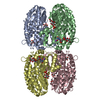 2pocSC  2putC 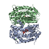 2puwC S: Starting model for refinement C: citing same article ( |
|---|---|
| Similar structure data |


-Links
 PDBj
PDBj


- Assembly
| Deposited unit | 
| ||||||||
|---|---|---|---|---|---|---|---|---|---|
| 1 |
| ||||||||
| Unit cell |
| ||||||||
| Details | The asymmetric unit contains the tetrameric biological assembly |
-Components
-Protein , 1 types, 4 molecules ABCD
| #1: Protein | Mass: 40614.738 Da / Num. of mol.: 4 / Fragment: isomerase domain Source method: isolated from a genetically manipulated source Source: (gene. exp.) Candida albicans (yeast) / Strain: SC5314 / Gene: GFA1 / Plasmid: pET23b-FRU / Production host:  References: UniProt: P53704, glutamine-fructose-6-phosphate transaminase (isomerizing) |
|---|
-Non-polymers , 5 types, 783 molecules 


| #2: Chemical |  Mass: 59.044 Da / Num. of mol.: 2 / Source method: obtained synthetically / Formula: C2H3O2 Mass: 59.044 Da / Num. of mol.: 2 / Source method: obtained synthetically / Formula: C2H3O2#3: Chemical | ChemComp-NA /  Mass: 22.990 Da / Num. of mol.: 4 / Source method: obtained synthetically / Formula: Na Mass: 22.990 Da / Num. of mol.: 4 / Source method: obtained synthetically / Formula: Na#4: Chemical | ChemComp-UD1 /  Mass: 607.354 Da / Num. of mol.: 4 / Source method: obtained synthetically / Formula: C17H27N3O17P2 Mass: 607.354 Da / Num. of mol.: 4 / Source method: obtained synthetically / Formula: C17H27N3O17P2#5: Chemical | ChemComp-M6R /  Mass: 261.167 Da / Num. of mol.: 4 / Source method: obtained synthetically / Formula: C6H16NO8P Mass: 261.167 Da / Num. of mol.: 4 / Source method: obtained synthetically / Formula: C6H16NO8P#6: Water | ChemComp-HOH / | Mass: 18.015 Da / Num. of mol.: 769 / Source method: isolated from a natural source / Formula: H2O |
|---|
-Experimental details
-Experiment
| Experiment | Method: X-RAY DIFFRACTION / Number of used crystals: 1 |
|---|
- Sample preparation
| Crystal | Density Matthews: 2.41 Å3/Da / Density % sol: 48.88 % |
|---|---|
| Crystal grow | Temperature: 277 K / Method: vapor diffusion, hanging drop / pH: 6.5 Details: 0.2 M magnesium acetate, 0.1 M sodium cacodylate pH 6.5, 30% v/v 2-methyl-2,4-pentanediol, 10-fold excess of UDP-GlcNAc and Glc-6-P; crystals soaked with large excess of 2-amino-2-deoxy-D- ...Details: 0.2 M magnesium acetate, 0.1 M sodium cacodylate pH 6.5, 30% v/v 2-methyl-2,4-pentanediol, 10-fold excess of UDP-GlcNAc and Glc-6-P; crystals soaked with large excess of 2-amino-2-deoxy-D-mannitol 6-phosphate, VAPOR DIFFUSION, HANGING DROP, temperature 277K |
-Data collection
| Diffraction | Mean temperature: 100 K |
|---|---|
| Diffraction source | Source: SYNCHROTRON / Site: EMBL/DESY, HAMBURG  / Beamline: X11 / Wavelength: 0.8162 Å / Beamline: X11 / Wavelength: 0.8162 Å |
| Detector | Type: MAR CCD 165 mm / Detector: CCD / Date: May 8, 2006 |
| Radiation | Monochromator: Si [111] / Protocol: SINGLE WAVELENGTH / Monochromatic (M) / Laue (L): M / Scattering type: x-ray |
| Radiation wavelength | Wavelength: 0.8162 Å / Relative weight: 1 |
| Reflection | Resolution: 1.9→20 Å / Num. all: 120934 / Num. obs: 120934 / % possible obs: 99.6 % / Observed criterion σ(F): 0 / Observed criterion σ(I): 0 / Redundancy: 4.1 % / Biso Wilson estimate: 23.2 Å2 / Rsym value: 0.058 / Net I/σ(I): 22 |
| Reflection shell | Resolution: 1.9→1.93 Å / Redundancy: 3.8 % / Mean I/σ(I) obs: 2.3 / Num. unique all: 5743 / Rsym value: 0.474 / % possible all: 94.7 |
- Processing
| Software |
| ||||||||||||||||||||||||||||||||||||||||||||||||||||||||||||||||||||||||||||||||||||||||||||||||||||||||||||||||||||||||||||||||||||||||||||||||||||||||||||||||||||||||||
|---|---|---|---|---|---|---|---|---|---|---|---|---|---|---|---|---|---|---|---|---|---|---|---|---|---|---|---|---|---|---|---|---|---|---|---|---|---|---|---|---|---|---|---|---|---|---|---|---|---|---|---|---|---|---|---|---|---|---|---|---|---|---|---|---|---|---|---|---|---|---|---|---|---|---|---|---|---|---|---|---|---|---|---|---|---|---|---|---|---|---|---|---|---|---|---|---|---|---|---|---|---|---|---|---|---|---|---|---|---|---|---|---|---|---|---|---|---|---|---|---|---|---|---|---|---|---|---|---|---|---|---|---|---|---|---|---|---|---|---|---|---|---|---|---|---|---|---|---|---|---|---|---|---|---|---|---|---|---|---|---|---|---|---|---|---|---|---|---|---|---|---|
| Refinement | Method to determine structure: MOLECULAR REPLACEMENT Starting model: 2POC Resolution: 1.9→19.93 Å / Cor.coef. Fo:Fc: 0.959 / Cor.coef. Fo:Fc free: 0.941 / SU B: 2.803 / SU ML: 0.085 / Isotropic thermal model: Isotropic / Cross valid method: THROUGHOUT / σ(F): 0 / σ(I): 0 / ESU R: 0.133 / ESU R Free: 0.128 / Stereochemistry target values: MAXIMUM LIKELIHOOD
| ||||||||||||||||||||||||||||||||||||||||||||||||||||||||||||||||||||||||||||||||||||||||||||||||||||||||||||||||||||||||||||||||||||||||||||||||||||||||||||||||||||||||||
| Solvent computation | Ion probe radii: 0.8 Å / Shrinkage radii: 0.8 Å / VDW probe radii: 1.2 Å / Solvent model: MASK | ||||||||||||||||||||||||||||||||||||||||||||||||||||||||||||||||||||||||||||||||||||||||||||||||||||||||||||||||||||||||||||||||||||||||||||||||||||||||||||||||||||||||||
| Displacement parameters | Biso mean: 25.539 Å2
| ||||||||||||||||||||||||||||||||||||||||||||||||||||||||||||||||||||||||||||||||||||||||||||||||||||||||||||||||||||||||||||||||||||||||||||||||||||||||||||||||||||||||||
| Refine analyze | Luzzati sigma a obs: 0.0845 Å | ||||||||||||||||||||||||||||||||||||||||||||||||||||||||||||||||||||||||||||||||||||||||||||||||||||||||||||||||||||||||||||||||||||||||||||||||||||||||||||||||||||||||||
| Refinement step | Cycle: LAST / Resolution: 1.9→19.93 Å
| ||||||||||||||||||||||||||||||||||||||||||||||||||||||||||||||||||||||||||||||||||||||||||||||||||||||||||||||||||||||||||||||||||||||||||||||||||||||||||||||||||||||||||
| Refine LS restraints |
| ||||||||||||||||||||||||||||||||||||||||||||||||||||||||||||||||||||||||||||||||||||||||||||||||||||||||||||||||||||||||||||||||||||||||||||||||||||||||||||||||||||||||||
| LS refinement shell | Resolution: 1.9→1.949 Å / Total num. of bins used: 20
|
