 Movie
Movie Controller

[English] 日本語
 Yorodumi
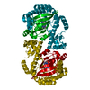
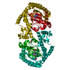
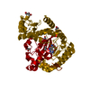
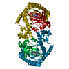
- PDB-2psg: REFINED STRUCTURE OF PORCINE PEPSINOGEN AT 1.8 ANGSTROMS RESOLUTION -
+ Open data

- Basic information
| Entry | Database: PDB / ID: 2psg | ||||||
|---|---|---|---|---|---|---|---|
| Title | REFINED STRUCTURE OF PORCINE PEPSINOGEN AT 1.8 ANGSTROMS RESOLUTION | ||||||
 Components Components | PEPSINOGEN | ||||||
 Keywords Keywords | HYDROLASE(ACID PROTEINASE ZYMOGEN) | ||||||
| Function / homology |  Function and homology information Function and homology informationSurfactant metabolism / pepsin A / digestion / aspartic-type endopeptidase activity / proteolysis / extracellular region Similarity search - Function | ||||||
| Biological species |  | ||||||
| Method |  X-RAY DIFFRACTION / Resolution: 1.8 Å X-RAY DIFFRACTION / Resolution: 1.8 Å | ||||||
 Authors Authors | James, M.N.G. / Sielecki, A.R. | ||||||
 Citation Citation | Journal: J.Mol.Biol. / Year: 1991 Title: Refined structure of porcine pepsinogen at 1.8 A resolution. Authors: Sielecki, A.R. / Fujinaga, M. / Read, R.J. / James, M.N. #1: Journal: Nature / Year: 1986Title: Molecular Structure of an Aspartic Proteinase Zymogen, Porcine Pepsinogen, at 1.8 Angstroms Resolution Authors: James, M.N.G. / Sielecki, A.R. #2: Journal: Biochemistry / Year: 1986Title: Characterization of Phosphoserine of Pepsinogen Using 31P Nuclear Magnetic Resonance: Corroboration of X-Ray Crystallographic Results Authors: Williams, S.P. / Bridger, W.A. / James, M.N.G. | ||||||
| History |
|
- Structure visualization
| Structure viewer | Molecule: MolmilJmol/JSmol |
|---|
- Downloads & links
-Download
| PDBx/mmCIF format | 2psg.cif.gz | 88.4 KB | Display | PDBx/mmCIF format |
|---|---|---|---|---|
| PDB format | pdb2psg.ent.gz | 65.4 KB | Display | PDB format |
| PDBx/mmJSON format | 2psg.json.gz | Tree view | PDBx/mmJSON format | |
| Others |  Other downloads Other downloads |
-Validation report
| Arichive directory | https://data.pdbj.org/pub/pdb/validation_reports/ps/2psgftp://data.pdbj.org/pub/pdb/validation_reports/ps/2psg | HTTPS FTP |
|---|
-Related structure data
| Similar structure data |
|---|


-Links
 PDBj
PDBj
- Assembly
| Deposited unit | 
| ||||||||
|---|---|---|---|---|---|---|---|---|---|
| 1 | 
| ||||||||
| Unit cell |
| ||||||||
| Atom site foot note | 1: RESIDUE PRO 23 IS A CIS PROLINE. 2: RESIDUE SER 68 IS PHOSHORYLATED. THE PHOSPHATE GROUP IS PRESENTED AS HET GROUP PO3 ON HET RECORDS AT THE END OF THE CHAIN. | ||||||||
| Components on special symmetry positions |
|
-Components
| #1: Protein | Mass: 39631.504 Da / Num. of mol.: 1 Source method: isolated from a genetically manipulated source Source: (gene. exp.) |
|---|---|
| #2: Water | ChemComp-HOH /  Mass: 18.015 Da / Num. of mol.: 237 / Source method: isolated from a natural source / Formula: H2O Mass: 18.015 Da / Num. of mol.: 237 / Source method: isolated from a natural source / Formula: H2O |
| Has protein modification | Y |
| Sequence details | THE SEQUENCE USED IN THIS ENTRY IS THAT OF P.SEPULVEDA, J.MARCINISZYN JR.,D.LIU,J.TANG (J.BIO.CHEM. ...THE SEQUENCE USED IN THIS ENTRY IS THAT OF P.SEPULVEDA, J.MARCINISZY |
-Experimental details
-Experiment
| Experiment | Method: X-RAY DIFFRACTION |
|---|
- Sample preparation
| Crystal | Density Matthews: 2.57 Å3/Da / Density % sol: 52.05 % | ||||||||||||||||||||||||
|---|---|---|---|---|---|---|---|---|---|---|---|---|---|---|---|---|---|---|---|---|---|---|---|---|---|
| Crystal grow | *PLUS pH: 6.1 / Method: vapor diffusion, hanging drop / Details: took James & Sielecki, 1986 from original paper | ||||||||||||||||||||||||
| Components of the solutions | *PLUS
|
-Data collection
| Radiation | Scattering type: x-ray |
|---|---|
| Radiation wavelength | Relative weight: 1 |
| Reflection | *PLUS Highest resolution: 1.8 Å / Lowest resolution: 40 Å / Num. obs: 27251 / Observed criterion σ(I): 2 / Num. measured all: 37575 / Rmerge(I) obs: 0.058 |
- Processing
| Software | Name: PROLSQ / Classification: refinement | |||||||||||||||||||||||||||||||||||||||||||||||||||||||||||||||
|---|---|---|---|---|---|---|---|---|---|---|---|---|---|---|---|---|---|---|---|---|---|---|---|---|---|---|---|---|---|---|---|---|---|---|---|---|---|---|---|---|---|---|---|---|---|---|---|---|---|---|---|---|---|---|---|---|---|---|---|---|---|---|---|---|
| Refinement | Resolution: 1.8→8 Å / σ(I): 1 /
| |||||||||||||||||||||||||||||||||||||||||||||||||||||||||||||||
| Refinement step | Cycle: LAST / Resolution: 1.8→8 Å
| |||||||||||||||||||||||||||||||||||||||||||||||||||||||||||||||
| Refine LS restraints |
| |||||||||||||||||||||||||||||||||||||||||||||||||||||||||||||||
| Software | *PLUS Name: PROLSQ / Classification: refinement | |||||||||||||||||||||||||||||||||||||||||||||||||||||||||||||||
| Refinement | *PLUS Rfactor obs: 0.164 | |||||||||||||||||||||||||||||||||||||||||||||||||||||||||||||||
| Solvent computation | *PLUS | |||||||||||||||||||||||||||||||||||||||||||||||||||||||||||||||
| Displacement parameters | *PLUS Biso mean: 15 Å2 |
