 Movie
Movie Controller

[English] 日本語
 Yorodumi
- PDB-2pqn: Crystal structure of yeast Fis1 complexed with a fragment of yeas... -
+ Open data

- Basic information
| Entry | Database: PDB / ID: 2pqn | ||||||
|---|---|---|---|---|---|---|---|
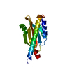
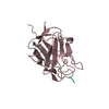
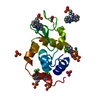
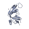
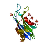
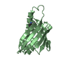
| Title | Crystal structure of yeast Fis1 complexed with a fragment of yeast Mdv1 | ||||||
 Components Components |
| ||||||
 Keywords Keywords | APOPTOSIS / TPR domain / protein-protein complex | ||||||
| Function / homology |  Function and homology information Function and homology informationClass I peroxisomal membrane protein import / peroxisome organization / peroxisome fission / mitochondrial fission / peroxisomal membrane / positive regulation of mitochondrial fission / ubiquitin binding / peroxisome / molecular adaptor activity / mitochondrial outer membrane ...Class I peroxisomal membrane protein import / peroxisome organization / peroxisome fission / mitochondrial fission / peroxisomal membrane / positive regulation of mitochondrial fission / ubiquitin binding / peroxisome / molecular adaptor activity / mitochondrial outer membrane / apoptotic process / lipid binding / mitochondrion Similarity search - Function | ||||||
| Biological species |  | ||||||
| Method |  X-RAY DIFFRACTION / SYNCHROTRON / MOLECULAR REPLACEMENT / Resolution: 2.15 Å X-RAY DIFFRACTION / SYNCHROTRON / MOLECULAR REPLACEMENT / Resolution: 2.15 Å | ||||||
 Authors Authors | Zhang, Y. / Chan, D.C. | ||||||
 Citation Citation | Journal: Proc.Natl.Acad.Sci.USA / Year: 2007 Title: Structural basis for recruitment of mitochondrial fission complexes by Fis1. Authors: Zhang, Y. / Chan, D.C. | ||||||
| History |
|
- Structure visualization
| Structure viewer | Molecule: MolmilJmol/JSmol |
|---|
- Downloads & links
-Download
| PDBx/mmCIF format | 2pqn.cif.gz | 44.9 KB | Display | PDBx/mmCIF format |
|---|---|---|---|---|
| PDB format | pdb2pqn.ent.gz | 30.8 KB | Display | PDB format |
| PDBx/mmJSON format | 2pqn.json.gz | Tree view | PDBx/mmJSON format | |
| Others |  Other downloads Other downloads |
-Validation report
| Arichive directory | https://data.pdbj.org/pub/pdb/validation_reports/pq/2pqnftp://data.pdbj.org/pub/pdb/validation_reports/pq/2pqn | HTTPS FTP |
|---|
-Related structure data
| Related structure data | 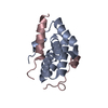 2pqrC 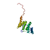 1y8mS C: citing same article ( S: Starting model for refinement |
|---|---|
| Similar structure data |

-Links
 PDBj
PDBj- Assembly
| Deposited unit | 
| ||||||||
|---|---|---|---|---|---|---|---|---|---|
| 1 |
| ||||||||
| Unit cell |
|
-Components
| #1: Protein | Mass: 15128.260 Da / Num. of mol.: 1 / Fragment: cytosolic portion Source method: isolated from a genetically manipulated source Source: (gene. exp.) Gene: FIS1, MDV2 / Plasmid: pBB75 / Production host:  |
|---|---|
| #2: Protein | Mass: 6037.681 Da / Num. of mol.: 1 / Fragment: N-terminal domain Source method: isolated from a genetically manipulated source Source: (gene. exp.) Gene: MDV1, FIS2, GAG3, NET2 / Plasmid: pET15b / Production host: |
| #3: Water | ChemComp-HOH /  Mass: 18.015 Da / Num. of mol.: 41 / Source method: isolated from a natural source / Formula: H2O Mass: 18.015 Da / Num. of mol.: 41 / Source method: isolated from a natural source / Formula: H2O |
-Experimental details
-Experiment
| Experiment | Method: X-RAY DIFFRACTION / Number of used crystals: 1 |
|---|
- Sample preparation
| Crystal | Density Matthews: 2.05 Å3/Da / Density % sol: 40.09 % |
|---|---|
| Crystal grow | Temperature: 295 K / Method: vapor diffusion, hanging drop / pH: 8 Details: 20% PEG3350, 0.1M Tris, pH 8.0, VAPOR DIFFUSION, HANGING DROP, temperature 295K |
-Data collection
| Diffraction | Mean temperature: 100 K |
|---|---|
| Diffraction source | Source: SYNCHROTRON / Site: SSRL  / Beamline: BL9-2 / Wavelength: 0.97946 / Beamline: BL9-2 / Wavelength: 0.97946 |
| Detector | Type: MARMOSAIC 325 mm CCD / Detector: CCD / Date: Apr 12, 2006 Details: Flat collimating mirror, double crystal monochromator, toroid focusing mirror |
| Radiation | Monochromator: Double crystal monochromator, Si(111) / Protocol: SINGLE WAVELENGTH / Monochromatic (M) / Laue (L): M / Scattering type: x-ray |
| Radiation wavelength | Wavelength: 0.97946 Å / Relative weight: 1 |
| Reflection | Resolution: 2.15→50 Å / Num. obs: 9834 / % possible obs: 98.6 % / Redundancy: 5.7 % / Rmerge(I) obs: 0.063 / Net I/σ(I): 13 |
| Reflection shell | Resolution: 2.15→2.23 Å / Redundancy: 4.3 % / Rmerge(I) obs: 0.359 / % possible all: 89.5 |
-Phasing
| Phasing MR | Method rotation: fast direct / Method translation: STRIPtrans_method |
|---|
- Processing
| Software |
| ||||||||||||||||||||||||||||||||||||||||||||||||||||||||||||||||||||||||||||||||||||||||||||||||||||||||||||||||||||||||||||||||||||||||||||||||||||||||||||||||||||||||||
|---|---|---|---|---|---|---|---|---|---|---|---|---|---|---|---|---|---|---|---|---|---|---|---|---|---|---|---|---|---|---|---|---|---|---|---|---|---|---|---|---|---|---|---|---|---|---|---|---|---|---|---|---|---|---|---|---|---|---|---|---|---|---|---|---|---|---|---|---|---|---|---|---|---|---|---|---|---|---|---|---|---|---|---|---|---|---|---|---|---|---|---|---|---|---|---|---|---|---|---|---|---|---|---|---|---|---|---|---|---|---|---|---|---|---|---|---|---|---|---|---|---|---|---|---|---|---|---|---|---|---|---|---|---|---|---|---|---|---|---|---|---|---|---|---|---|---|---|---|---|---|---|---|---|---|---|---|---|---|---|---|---|---|---|---|---|---|---|---|---|---|---|
| Refinement | Method to determine structure: MOLECULAR REPLACEMENT Starting model: PDB entry 1Y8M Resolution: 2.15→29.81 Å / Cor.coef. Fo:Fc: 0.953 / Cor.coef. Fo:Fc free: 0.939 / SU B: 11.018 / SU ML: 0.147 / TLS residual ADP flag: LIKELY RESIDUAL / Cross valid method: THROUGHOUT / ESU R: 0.254 / ESU R Free: 0.197 / Stereochemistry target values: MAXIMUM LIKELIHOOD / Details: HYDROGENS HAVE BEEN ADDED IN THE RIDING POSITIONS
| ||||||||||||||||||||||||||||||||||||||||||||||||||||||||||||||||||||||||||||||||||||||||||||||||||||||||||||||||||||||||||||||||||||||||||||||||||||||||||||||||||||||||||
| Solvent computation | Ion probe radii: 0.8 Å / Shrinkage radii: 0.8 Å / VDW probe radii: 1.2 Å / Solvent model: MASK | ||||||||||||||||||||||||||||||||||||||||||||||||||||||||||||||||||||||||||||||||||||||||||||||||||||||||||||||||||||||||||||||||||||||||||||||||||||||||||||||||||||||||||
| Displacement parameters | Biso mean: 43.17 Å2
| ||||||||||||||||||||||||||||||||||||||||||||||||||||||||||||||||||||||||||||||||||||||||||||||||||||||||||||||||||||||||||||||||||||||||||||||||||||||||||||||||||||||||||
| Refinement step | Cycle: LAST / Resolution: 2.15→29.81 Å
| ||||||||||||||||||||||||||||||||||||||||||||||||||||||||||||||||||||||||||||||||||||||||||||||||||||||||||||||||||||||||||||||||||||||||||||||||||||||||||||||||||||||||||
| Refine LS restraints |
| ||||||||||||||||||||||||||||||||||||||||||||||||||||||||||||||||||||||||||||||||||||||||||||||||||||||||||||||||||||||||||||||||||||||||||||||||||||||||||||||||||||||||||
| LS refinement shell | Resolution: 2.147→2.203 Å / Total num. of bins used: 20
| ||||||||||||||||||||||||||||||||||||||||||||||||||||||||||||||||||||||||||||||||||||||||||||||||||||||||||||||||||||||||||||||||||||||||||||||||||||||||||||||||||||||||||
| Refinement TLS params. | Method: refined / Refine-ID: X-RAY DIFFRACTION
| ||||||||||||||||||||||||||||||||||||||||||||||||||||||||||||||||||||||||||||||||||||||||||||||||||||||||||||||||||||||||||||||||||||||||||||||||||||||||||||||||||||||||||
| Refinement TLS group | Refine-ID: X-RAY DIFFRACTION / Selection: ALL
|
