 Movie
Movie Controller

[English] 日本語
 Yorodumi
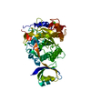
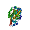
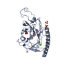
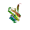
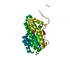
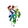
- PDB-2pqg: Crystal structure of inactive ribosome inactivating protein from ... -
+ Open data

- Basic information
| Entry | Database: PDB / ID: 2pqg | ||||||
|---|---|---|---|---|---|---|---|
| Title | Crystal structure of inactive ribosome inactivating protein from maize (b-32) | ||||||
 Components Components | Ribosome-inactivating protein 3 | ||||||
 Keywords Keywords | HYDROLASE / Pro-RIP / ribosome inactivating protein / maize | ||||||
| Function / homology |  Function and homology information Function and homology informationrRNA N-glycosylase / rRNA N-glycosylase activity / defense response / toxin activity / negative regulation of translation / cytoplasm Similarity search - Function | ||||||
| Biological species |  | ||||||
| Method |  X-RAY DIFFRACTION / SYNCHROTRON / MOLECULAR REPLACEMENT / Resolution: 2.38 Å X-RAY DIFFRACTION / SYNCHROTRON / MOLECULAR REPLACEMENT / Resolution: 2.38 Å | ||||||
 Authors Authors | Mak, A.N.S. / Wong, Y.T. / Young, J.A. / Cha, S.S. / Sze, K.H. / Au, S.W.N. / Wong, K.B. / Shaw, P.C. | ||||||
 Citation Citation | Journal: Nucleic Acids Res. / Year: 2007 Title: Structure-function study of maize ribosome-inactivating protein: implications for the internal inactivation region and the sole glutamate in the active site. Authors: Mak, A.N. / Wong, Y.T. / An, Y.J. / Cha, S.S. / Sze, K.H. / Au, S.W. / Wong, K.B. / Shaw, P.C. #1: Journal: J.Biol.Chem. / Year: 1991Title: Characterization and molecular cloning of a proenzyme form of a ribosome-inactivating protein from maize. Novel mechanism of proenzyme activation by proteolytic removal of a 2.8-kilodalton internal peptide segment Authors: Walsh, T.A. / Morgan, A.E. / Hey, T.D. | ||||||
| History |
|
- Structure visualization
| Structure viewer | Molecule: MolmilJmol/JSmol |
|---|
- Downloads & links
-Download
| PDBx/mmCIF format | 2pqg.cif.gz | 111.6 KB | Display | PDBx/mmCIF format |
|---|---|---|---|---|
| PDB format | pdb2pqg.ent.gz | 86.1 KB | Display | PDB format |
| PDBx/mmJSON format | 2pqg.json.gz | Tree view | PDBx/mmJSON format | |
| Others |  Other downloads Other downloads |
-Validation report
| Arichive directory | https://data.pdbj.org/pub/pdb/validation_reports/pq/2pqgftp://data.pdbj.org/pub/pdb/validation_reports/pq/2pqg | HTTPS FTP |
|---|
-Related structure data
| Related structure data |  2pqiC  2pqjC 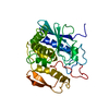 1rtcS S: Starting model for refinement C: citing same article ( |
|---|---|
| Similar structure data |


-Links
 PDBj
PDBj

- Assembly
| Deposited unit | 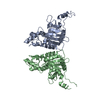
| ||||||||
|---|---|---|---|---|---|---|---|---|---|
| 1 | 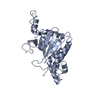
| ||||||||
| 2 | 
| ||||||||
| Unit cell |
|
-Components
| #1: Protein | Mass: 29607.010 Da / Num. of mol.: 2 Source method: isolated from a genetically manipulated source Source: (gene. exp.)  #2: Water | ChemComp-HOH / |  Mass: 18.015 Da / Num. of mol.: 134 / Source method: isolated from a natural source / Formula: H2O Mass: 18.015 Da / Num. of mol.: 134 / Source method: isolated from a natural source / Formula: H2O |
|---|
-Experimental details
-Experiment
| Experiment | Method: X-RAY DIFFRACTION / Number of used crystals: 1 |
|---|
- Sample preparation
| Crystal | Density Matthews: 2.4 Å3/Da / Density % sol: 48.71 % |
|---|---|
| Crystal grow | Temperature: 289 K / Method: vapor diffusion, sitting drop / pH: 3.5 Details: 2M ammonium sulphate, 0.25 M Tris-HCl and 0.1 M sodium acetate tri-hydrate, pH 3.5., VAPOR DIFFUSION, SITTING DROP, temperature 289K |
-Data collection
| Diffraction | Mean temperature: 100 K |
|---|---|
| Diffraction source | Source: SYNCHROTRON / Site: PAL/PLS  / Beamline: 6B / Wavelength: 1.5418 Å / Beamline: 6B / Wavelength: 1.5418 Å |
| Detector | Type: MAC Science DIP-2030 / Detector: IMAGE PLATE / Date: Nov 20, 2006 |
| Radiation | Protocol: SINGLE WAVELENGTH / Monochromatic (M) / Laue (L): M / Scattering type: x-ray |
| Radiation wavelength | Wavelength: 1.5418 Å / Relative weight: 1 |
| Reflection | Resolution: 2.38→30 Å / Num. all: 55918 / Num. obs: 19282 / % possible obs: 84.1 % / Observed criterion σ(F): 0 / Observed criterion σ(I): 0 / Redundancy: 2.9 % / Rmerge(I) obs: 0.118 / Net I/σ(I): 7.1 |
| Reflection shell | Resolution: 2.38→2.48 Å / Redundancy: 2.1 % / Rmerge(I) obs: 0.342 / Mean I/σ(I) obs: 1.6 / % possible all: 70.6 |
- Processing
| Software |
| |||||||||||||||||||||||||
|---|---|---|---|---|---|---|---|---|---|---|---|---|---|---|---|---|---|---|---|---|---|---|---|---|---|---|
| Refinement | Method to determine structure: MOLECULAR REPLACEMENT Starting model: 1RTC Resolution: 2.38→30 Å / Cross valid method: THROUGHOUT / σ(F): 0 / σ(I): 0 / Stereochemistry target values: Engh & Huber
| |||||||||||||||||||||||||
| Refinement step | Cycle: LAST / Resolution: 2.38→30 Å
| |||||||||||||||||||||||||
| Refine LS restraints |
| |||||||||||||||||||||||||
| LS refinement shell | Highest resolution: 2.38 Å
|
