- PDB-2pfu: NMR structure determination of the periplasmic domain of ExbD fro... -
+
Open data
ID or keywords:
Loading...
-
Basic information
Entry
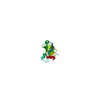
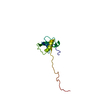
Database: PDB / ID: 2pfu
Title
NMR structure determination of the periplasmic domain of ExbD from E.coli
Components
Biopolymer transport exbD protein
Keywords
TRANSPORT PROTEIN / ExbD / TonB system / proton motive force / periplasmic domain
Function / homology
Function and homology information
energy transducer activity / Metal ion assimilation from the host / bacteriocin transport / cobalamin transport / intracellular monoatomic cation homeostasis / Iron assimilation using enterobactin / plasma membrane protein complex / transmembrane transporter complex / Heme assimilation / transmembrane transporter activity ...energy transducer activity / Metal ion assimilation from the host / bacteriocin transport / cobalamin transport / intracellular monoatomic cation homeostasis / Iron assimilation using enterobactin / plasma membrane protein complex / transmembrane transporter complex / Heme assimilation / transmembrane transporter activity / cell outer membrane / protein transport / intracellular iron ion homeostasis / protein stabilization / protein homodimerization activity / membrane / identical protein binding / plasma membrane Similarity search - Function
Nucleotidyltransferase; domain 5 - #270 / TonB system transport protein ExbD type-1 / Biopolymer transport protein ExbD/TolR / Biopolymer transport protein ExbD/TolR / Nucleotidyltransferase; domain 5 / 2-Layer Sandwich / Alpha Beta Similarity search - Domain/homology
In the structure databanks used in Yorodumi, some data are registered as the other names, "COVID-19 virus" and "2019-nCoV". Here are the details of the virus and the list of structure data.
Jan 31, 2019. EMDB accession codes are about to change! (news from PDBe EMDB page)
EMDB accession codes are about to change! (news from PDBe EMDB page)
The allocation of 4 digits for EMDB accession codes will soon come to an end. Whilst these codes will remain in use, new EMDB accession codes will include an additional digit and will expand incrementally as the available range of codes is exhausted. The current 4-digit format prefixed with “EMD-” (i.e. EMD-XXXX) will advance to a 5-digit format (i.e. EMD-XXXXX), and so on. It is currently estimated that the 4-digit codes will be depleted around Spring 2019, at which point the 5-digit format will come into force.
The EM Navigator/Yorodumi systems omit the EMD- prefix.
Related info.:Q: What is EMD? / ID/Accession-code notation in Yorodumi/EM Navigator
Yorodumi is a browser for structure data from EMDB, PDB, SASBDB, etc.
This page is also the successor to EM Navigator detail page, and also detail information page/front-end page for Omokage search.
The word "yorodu" (or yorozu) is an old Japanese word meaning "ten thousand". "mi" (miru) is to see.
Related info.:EMDB / PDB / SASBDB / Comparison of 3 databanks / Yorodumi Search / Aug 31, 2016. New EM Navigator & Yorodumi / Yorodumi Papers / Jmol/JSmol / Function and homology information / Changes in new EM Navigator and Yorodumi
 Movie
Movie Controller

 Yorodumi
 Open data
 Basic information
 Components
 Keywords
 Function and homology information
 Authors
 Citation
 Structure visualization
 Downloads & links
 Other downloads
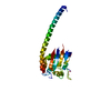


 PDBj
PDBj Assembly
 Sample preparation
 Processing
 CYANA
CYANA