 Movie
Movie Controller

[English] 日本語
 Yorodumi
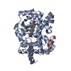
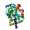
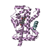
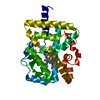
- PDB-2p7o: Crystal structure of genomically encoded fosfomycin resistance pr... -
+ Open data

- Basic information
| Entry | Database: PDB / ID: 2p7o | ||||||
|---|---|---|---|---|---|---|---|
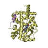
| Title | Crystal structure of genomically encoded fosfomycin resistance protein, FosX, from Listeria monocytogenes (tetragonal form) | ||||||
 Components Components | Glyoxalase family protein | ||||||
 Keywords Keywords | METAL BINDING PROTEIN / hydrolase / FOSFOMYCIN RESISTANCE PROTEIN / MN BINDING / ANTIBIOTIC RESISTANCE | ||||||
| Function / homology |  Function and homology information Function and homology information | ||||||
| Biological species |  Listeria monocytogenes (bacteria) Listeria monocytogenes (bacteria) | ||||||
| Method |  X-RAY DIFFRACTION / MOLECULAR REPLACEMENT / Resolution: 1.44 Å X-RAY DIFFRACTION / MOLECULAR REPLACEMENT / Resolution: 1.44 Å | ||||||
 Authors Authors | Fillgrove, K.L. / Pakhomova, S. / Schaab, M. / Newcomer, M.E. / Armstrong, R.N. | ||||||
 Citation Citation | Journal: Biochemistry / Year: 2007 Title: Structure and Mechanism of the Genomically Encoded Fosfomycin Resistance Protein, FosX, from Listeria monocytogenes. Authors: Fillgrove, K.L. / Pakhomova, S. / Schaab, M.R. / Newcomer, M.E. / Armstrong, R.N. | ||||||
| History |
|
- Structure visualization
| Structure viewer | Molecule: MolmilJmol/JSmol |
|---|
- Downloads & links
-Download
| PDBx/mmCIF format | 2p7o.cif.gz | 125.4 KB | Display | PDBx/mmCIF format |
|---|---|---|---|---|
| PDB format | pdb2p7o.ent.gz | 96.9 KB | Display | PDB format |
| PDBx/mmJSON format | 2p7o.json.gz | Tree view | PDBx/mmJSON format | |
| Others |  Other downloads Other downloads |
-Validation report
| Arichive directory | https://data.pdbj.org/pub/pdb/validation_reports/p7/2p7oftp://data.pdbj.org/pub/pdb/validation_reports/p7/2p7o | HTTPS FTP |
|---|
-Related structure data
| Related structure data |  2p7kSC 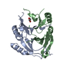 2p7lC  2p7mC  2p7pC 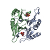 2p7qC S: Starting model for refinement C: citing same article ( |
|---|---|
| Similar structure data |


-Links
 PDBj
PDBj- Assembly
| Deposited unit | 
| ||||||||
|---|---|---|---|---|---|---|---|---|---|
| 1 |
| ||||||||
| Unit cell |
|
-Components
| #1: Protein | Mass: 15590.603 Da / Num. of mol.: 2 Source method: isolated from a genetically manipulated source Source: (gene. exp.) Listeria monocytogenes (bacteria) / Strain: EGD-e / Plasmid: pET20b(+) / Production host: #2: Chemical |   Mass: 54.938 Da / Num. of mol.: 2 / Source method: obtained synthetically / Formula: Mn Mass: 54.938 Da / Num. of mol.: 2 / Source method: obtained synthetically / Formula: Mn#3: Water | ChemComp-HOH / |  Mass: 18.015 Da / Num. of mol.: 168 / Source method: isolated from a natural source / Formula: H2O Mass: 18.015 Da / Num. of mol.: 168 / Source method: isolated from a natural source / Formula: H2O |
|---|
-Experimental details
-Experiment
| Experiment | Method: X-RAY DIFFRACTION |
|---|
- Sample preparation
| Crystal | Density Matthews: 1.99 Å3/Da / Density % sol: 42.6 % |
|---|---|
| Crystal grow | Temperature: 295 K / Method: vapor diffusion, hanging drop / pH: 8.1 Details: 22% PEG 3350, 0.1 M Tris-HCl, 0.2 M MgCl2, 5% isopropanol, pH 8.1, VAPOR DIFFUSION, HANGING DROP, temperature 295K |
-Data collection
| Diffraction | Mean temperature: 100 K |
|---|---|
| Diffraction source | Source: ROTATING ANODE / Type: ENRAF-NONIUS FR591 / Wavelength: 1.5418 Å |
| Detector | Type: MAR scanner 345 mm plate / Detector: IMAGE PLATE / Date: Apr 14, 2002 / Details: mirrors |
| Radiation | Monochromator: mirrors / Protocol: SINGLE WAVELENGTH / Monochromatic (M) / Laue (L): M / Scattering type: x-ray |
| Radiation wavelength | Wavelength: 1.5418 Å / Relative weight: 1 |
| Reflection | Resolution: 1.44→30 Å / Num. all: 41281 / Num. obs: 41281 / % possible obs: 86.5 % / Observed criterion σ(I): -3 / Redundancy: 4.6 % / Rsym value: 0.048 / Net I/σ(I): 24.4 |
| Reflection shell | Resolution: 1.44→1.46 Å / Redundancy: 4.2 % / Mean I/σ(I) obs: 2.1 / Num. unique all: 2325 / Rsym value: 0.478 / % possible all: 97.6 |
- Processing
| Software |
| |||||||||||||||||||||||||||||||||
|---|---|---|---|---|---|---|---|---|---|---|---|---|---|---|---|---|---|---|---|---|---|---|---|---|---|---|---|---|---|---|---|---|---|---|
| Refinement | Method to determine structure: MOLECULAR REPLACEMENT Starting model: PDB ENTRY 2P7K Resolution: 1.44→10 Å / Num. parameters: 20274 / Num. restraintsaints: 25273 / Cross valid method: FREE R / σ(F): 0 / σ(I): 0 / Stereochemistry target values: ENGH AND HUBER / Details: ANISOTROPIC REFINEMENT
| |||||||||||||||||||||||||||||||||
| Refine analyze | Num. disordered residues: 7 / Occupancy sum hydrogen: 0 / Occupancy sum non hydrogen: 2223 | |||||||||||||||||||||||||||||||||
| Refinement step | Cycle: LAST / Resolution: 1.44→10 Å
| |||||||||||||||||||||||||||||||||
| Refine LS restraints |
|
