[English] 日本語
Yorodumi
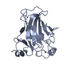
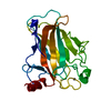
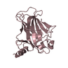
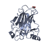
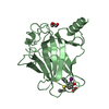
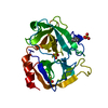
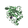
- PDB-2p52: mouse p53 DNA-binding domain in zinc-free oxidized state -

+
Open data


ID or keywords:

Loading...

-
Basic information

Entry
Database: PDB / ID: 2p52
Titlemouse p53 DNA-binding domain in zinc-free oxidized state
ComponentsCellular tumor antigen p53
KeywordsDNA BINDING PROTEIN / zinc finger / disulfide bond / oxidation immunoglobulin fold
Function / homology
Function and homology information


Formation of Senescence-Associated Heterochromatin Foci (SAHF) / Regulation of TP53 Expression / Regulation of TP53 Activity through Acetylation / Transcriptional activation of cell cycle inhibitor p21 / Regulation of TP53 Activity through Association with Co-factors / PI5P Regulates TP53 Acetylation / RUNX3 regulates CDKN1A transcription / DNA Damage/Telomere Stress Induced Senescence / G2/M DNA damage checkpoint / Stabilization of p53 ...Formation of Senescence-Associated Heterochromatin Foci (SAHF) / Regulation of TP53 Expression / Regulation of TP53 Activity through Acetylation / Transcriptional activation of cell cycle inhibitor p21 / Regulation of TP53 Activity through Association with Co-factors / PI5P Regulates TP53 Acetylation / RUNX3 regulates CDKN1A transcription / DNA Damage/Telomere Stress Induced Senescence / G2/M DNA damage checkpoint / Stabilization of p53 / Regulation of TP53 Activity through Methylation / Regulation of TP53 Degradation / Oncogene Induced Senescence / Autodegradation of the E3 ubiquitin ligase COP1 / G2/M Checkpoints / Ovarian tumor domain proteases / Recruitment and ATM-mediated phosphorylation of repair and signaling proteins at DNA double strand breaks / PKR-mediated signaling / The role of GTSE1 in G2/M progression after G2 checkpoint / Oxidative Stress Induced Senescence / Regulation of TP53 Activity through Phosphorylation / Ub-specific processing proteases / negative regulation of G1 to G0 transition / negative regulation of pentose-phosphate shunt / ATP-dependent DNA/DNA annealing activity / oligodendrocyte apoptotic process / positive regulation of thymocyte apoptotic process / oxidative stress-induced premature senescence / bone marrow development / cellular response to actinomycin D / circadian behavior / positive regulation of programmed necrotic cell death / mRNA transcription / hematopoietic stem cell differentiation / response to X-ray / entrainment of circadian clock by photoperiod / positive regulation of release of cytochrome c from mitochondria / negative regulation of telomere maintenance via telomerase / intrinsic apoptotic signaling pathway in response to DNA damage by p53 class mediator / TFIID-class transcription factor complex binding / replicative senescence / viral process / intrinsic apoptotic signaling pathway in response to endoplasmic reticulum stress / determination of adult lifespan / positive regulation of RNA polymerase II transcription preinitiation complex assembly / negative regulation of fibroblast proliferation / positive regulation of execution phase of apoptosis / type II interferon-mediated signaling pathway / core promoter sequence-specific DNA binding / mitotic G1 DNA damage checkpoint signaling / positive regulation of intrinsic apoptotic signaling pathway / MDM2/MDM4 family protein binding / DNA damage response, signal transduction by p53 class mediator / protein phosphatase 2A binding / transcription initiation-coupled chromatin remodeling / molecular function activator activity / tumor necrosis factor-mediated signaling pathway / cellular response to ionizing radiation / cellular response to xenobiotic stimulus / cellular response to gamma radiation / mRNA 3'-UTR binding / protein tetramerization / circadian rhythm / promoter-specific chromatin binding / molecular condensate scaffold activity / negative regulation of cell growth / nucleotide-excision repair / receptor tyrosine kinase binding / cellular senescence / PML body / positive regulation of miRNA transcription / DNA-binding transcription repressor activity, RNA polymerase II-specific / transcription coactivator binding / positive regulation of reactive oxygen species metabolic process / intracellular protein localization / histone deacetylase binding / autophagy / nuclear matrix / disordered domain specific binding / p53 binding / cellular response to UV / positive regulation of cellular senescence / transcription regulator complex / protein-folding chaperone binding / protease binding / DNA-binding transcription activator activity, RNA polymerase II-specific / cellular response to hypoxia / RNA polymerase II-specific DNA-binding transcription factor binding / regulation of apoptotic process / Ras protein signal transduction / DNA-binding transcription factor activity, RNA polymerase II-specific / positive regulation of apoptotic process / RNA polymerase II cis-regulatory region sequence-specific DNA binding / mitochondrial matrix / negative regulation of cell population proliferation / protein heterodimerization activity / copper ion binding / negative regulation of DNA-templated transcription / ubiquitin protein ligase binding / centrosome
Similarity search - Function
Immunoglobulin-like - #720 / p53 transactivation domain / P53 transactivation motif / : / p53 family signature. / p53, tetramerisation domain / P53 tetramerisation motif / p53, DNA-binding domain / P53 DNA-binding domain / p53 tumour suppressor family ...Immunoglobulin-like - #720 / p53 transactivation domain / P53 transactivation motif / : / p53 family signature. / p53, tetramerisation domain / P53 tetramerisation motif / p53, DNA-binding domain / P53 DNA-binding domain / p53 tumour suppressor family / p53-like tetramerisation domain superfamily / p53/RUNT-type transcription factor, DNA-binding domain superfamily / p53-like transcription factor, DNA-binding / Immunoglobulin-like / Sandwich / Mainly Beta
Similarity search - Domain/homology
Cellular tumor antigen p53
Similarity search - Component
Biological speciesMus musculus (house mouse)
MethodX-RAY DIFFRACTION / SYNCHROTRON / MOLECULAR REPLACEMENT / Resolution: 1.5 Å
AuthorsKwon, E. / Kim, D.Y. / Suh, S.W. / Kim, K.K.
CitationJournal: Proteins / Year: 2008
Title: Crystal structure of the mouse p53 core domain in zinc-free state.
Authors: Kwon, E. / Kim, D.Y. / Suh, S.W. / Kim, K.K.
History
DepositionMar 14, 2007Deposition site: RCSB / Processing site: PDBJ
Revision 1.0Jan 29, 2008Provider: repository / Type: Initial release
Revision 1.1Jul 13, 2011Group: Version format compliance
Revision 1.2Oct 25, 2023Group: Data collection / Database references / Refinement description
Category: chem_comp_atom / chem_comp_bond ...chem_comp_atom / chem_comp_bond / database_2 / pdbx_initial_refinement_model
Item: _database_2.pdbx_DOI / _database_2.pdbx_database_accession
Revision 1.3Oct 9, 2024Group: Structure summary / Category: pdbx_entry_details / pdbx_modification_feature

-
Structure visualization

Structure viewerMolecule:
MolmilJmol/JSmol

Downloads & links

-
Assembly

Deposited unit
A: Cellular tumor antigen p53


Theoretical massNumber of molelcules
Total (without water)22,2141
Polymers22,2141
Non-polymers00
Water5,783321
1


  • Idetical with deposited unit
  • defined by author
TypeNameSymmetry operationNumber
identity operation1_555x,y,z1
Unit cell
Length a, b, c (Å)45.235, 59.939, 70.462
Angle α, β, γ (deg.)90.00, 90.00, 90.00
Int Tables number19
Space group name H-MP212121

-
Components

#1: Protein Cellular tumor antigen p53 / Tumor suppressor p53


Mass: 22214.291 Da / Num. of mol.: 1 / Fragment: DNA-binding domain
Source method: isolated from a genetically manipulated source
Source: (gene. exp.) Mus musculus (house mouse) / Plasmid: pET22b+ / Species (production host): Escherichia coli / Production host: Escherichia coli BL21(DE3) (bacteria) / Strain (production host): BL21(DE3) / References: UniProt: P02340

#2: Water ChemComp-HOH / water


Mass: 18.015 Da / Num. of mol.: 321 / Source method: isolated from a natural source / Formula: H2O
Has protein modificationY

-
Experimental details

-
Experiment

ExperimentMethod: X-RAY DIFFRACTION / Number of used crystals: 1

-
Sample preparation

CrystalDensity Matthews: 2.15 Å3/Da / Density % sol: 42.75 %
Crystal growTemperature: 277 K / Method: microbatch / pH: 6 / Details: pH 6.0, microbatch, temperature 277K

-
Data collection

DiffractionMean temperature: 100 K
Diffraction sourceSource: SYNCHROTRON / Site: Photon Factory / Beamline: AR-NW12A / Wavelength: 1 Å
DetectorType: ADSC QUANTUM 210 / Detector: CCD
RadiationProtocol: SINGLE WAVELENGTH / Monochromatic (M) / Laue (L): M / Scattering type: x-ray
Radiation wavelengthWavelength: 1 Å / Relative weight: 1
ReflectionResolution: 1.5→20 Å / Num. obs: 60182 / % possible obs: 99.8 % / Redundancy: 6.5 % / Biso Wilson estimate: 14.7 Å2 / Rmerge(I) obs: 0.087 / Rsym value: 0.072 / Net I/σ(I): 13.4
Reflection shellResolution: 1.5→1.55 Å / Redundancy: 6.4 % / Rmerge(I) obs: 0.374 / % possible all: 99.9

-
Processing

Software
NameVersionClassification
CNS1.2refinement
HKL-2000data collection
HKL-2000data reduction
HKL-2000data scaling
EPMRphasing
RefinementMethod to determine structure: MOLECULAR REPLACEMENT
Starting model: 1HU8
Resolution: 1.5→19.69 Å / Rfactor Rfree error: 0.006 / Data cutoff high absF: 1610907.56 / Data cutoff low absF: 0 / Isotropic thermal model: RESTRAINED / Cross valid method: THROUGHOUT / σ(F): 0
RfactorNum. reflection% reflectionSelection details
Rfree0.236 1554 5 %RANDOM
Rwork0.211 ---
obs0.211 31294 99.6 %-
Solvent computationSolvent model: FLAT MODEL / Bsol: 43.355 Å2 / ksol: 0.35 e/Å3
Displacement parametersBiso mean: 17.5 Å2
Baniso -1Baniso -2Baniso -3
1-1.68 Å20 Å20 Å2
2--1.93 Å20 Å2
3----3.61 Å2
Refine analyze
FreeObs
Luzzati coordinate error0.2 Å0.18 Å
Luzzati d res low-5 Å
Luzzati sigma a0.07 Å0.06 Å
Refinement stepCycle: LAST / Resolution: 1.5→19.69 Å
ProteinNucleic acidLigandSolventTotal
Num. atoms1553 0 0 321 1874
Refine LS restraints
Refine-IDTypeDev idealDev ideal target
X-RAY DIFFRACTIONc_bond_d0.005
X-RAY DIFFRACTIONc_bond_d_na
X-RAY DIFFRACTIONc_bond_d_prot
X-RAY DIFFRACTIONc_angle_d
X-RAY DIFFRACTIONc_angle_d_na
X-RAY DIFFRACTIONc_angle_d_prot
X-RAY DIFFRACTIONc_angle_deg1.3
X-RAY DIFFRACTIONc_angle_deg_na
X-RAY DIFFRACTIONc_angle_deg_prot
X-RAY DIFFRACTIONc_dihedral_angle_d24.6
X-RAY DIFFRACTIONc_dihedral_angle_d_na
X-RAY DIFFRACTIONc_dihedral_angle_d_prot
X-RAY DIFFRACTIONc_improper_angle_d0.83
X-RAY DIFFRACTIONc_improper_angle_d_na
X-RAY DIFFRACTIONc_improper_angle_d_prot
X-RAY DIFFRACTIONc_mcbond_it1.181.5
X-RAY DIFFRACTIONc_mcangle_it1.782
X-RAY DIFFRACTIONc_scbond_it1.992
X-RAY DIFFRACTIONc_scangle_it2.772.5
LS refinement shellResolution: 1.5→1.59 Å / Rfactor Rfree error: 0.016 / Total num. of bins used: 6
RfactorNum. reflection% reflection
Rfree0.247 248 4.8 %
Rwork0.216 4878 -
obs--99.9 %
Xplor file
Refine-IDSerial noParam fileTopol file
X-RAY DIFFRACTION1protein_rep.paramprotein.top
X-RAY DIFFRACTION2water_rep.paramwater.top

+
About Yorodumi

-
News

-
Feb 9, 2022. New format data for meta-information of EMDB entries

New format data for meta-information of EMDB entries

  • Version 3 of the EMDB header file is now the official format.
  • The previous official version 1.9 will be removed from the archive.

Related info.:EMDB header

External links:wwPDB to switch to version 3 of the EMDB data model

-
Aug 12, 2020. Covid-19 info

Covid-19 info

URL: https://pdbj.org/emnavi/covid19.php

New page: Covid-19 featured information page in EM Navigator.

Related info.:Covid-19 info / Mar 5, 2020. Novel coronavirus structure data

+
Mar 5, 2020. Novel coronavirus structure data

Novel coronavirus structure data

Related info.:Yorodumi Speices / Aug 12, 2020. Covid-19 info

External links:COVID-19 featured content - PDBj / Molecule of the Month (242):Coronavirus Proteases

+
Jan 31, 2019. EMDB accession codes are about to change! (news from PDBe EMDB page)

EMDB accession codes are about to change! (news from PDBe EMDB page)

  • The allocation of 4 digits for EMDB accession codes will soon come to an end. Whilst these codes will remain in use, new EMDB accession codes will include an additional digit and will expand incrementally as the available range of codes is exhausted. The current 4-digit format prefixed with “EMD-” (i.e. EMD-XXXX) will advance to a 5-digit format (i.e. EMD-XXXXX), and so on. It is currently estimated that the 4-digit codes will be depleted around Spring 2019, at which point the 5-digit format will come into force.
  • The EM Navigator/Yorodumi systems omit the EMD- prefix.

Related info.:Q: What is EMD? / ID/Accession-code notation in Yorodumi/EM Navigator

External links:EMDB Accession Codes are Changing Soon! / Contact to PDBj

+
Jul 12, 2017. Major update of PDB

Major update of PDB

  • wwPDB released updated PDB data conforming to the new PDBx/mmCIF dictionary.
  • This is a major update changing the version number from 4 to 5, and with Remediation, in which all the entries are updated.
  • In this update, many items about electron microscopy experimental information are reorganized (e.g. em_software).
  • Now, EM Navigator and Yorodumi are based on the updated data.

External links:wwPDB Remediation / Enriched Model Files Conforming to OneDep Data Standards Now Available in the PDB FTP Archive

-
Yorodumi

Thousand views of thousand structures

  • Yorodumi is a browser for structure data from EMDB, PDB, SASBDB, etc.
  • This page is also the successor to EM Navigator detail page, and also detail information page/front-end page for Omokage search.
  • The word "yorodu" (or yorozu) is an old Japanese word meaning "ten thousand". "mi" (miru) is to see.

Related info.:EMDB / PDB / SASBDB / Comparison of 3 databanks / Yorodumi Search / Aug 31, 2016. New EM Navigator & Yorodumi / Yorodumi Papers / Jmol/JSmol / Function and homology information / Changes in new EM Navigator and Yorodumi

Read more