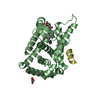
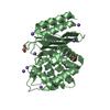
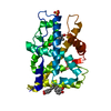
- PDB-2oz5: Crystal structure of Mycobacterium tuberculosis protein tyrosine ... -
+
Open data
ID or keywords:
Loading...
-
Basic information
Entry
Database: PDB / ID: 2oz5
Title
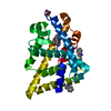
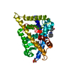
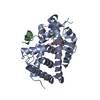
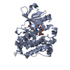
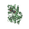
Crystal structure of Mycobacterium tuberculosis protein tyrosine phosphatase PtpB in complex with the specific inhibitor OMTS
Components
Phosphotyrosine protein phosphatase ptpb
Keywords
STRUCTURAL GENOMICS / UNKNOWN FUNCTION / Protein tyrosine phosphatase in complex with small molecule inhibitor / TB Structural Genomics Consortium / TBSGC
Function / homology
Function and homology information
phosphatidylinositol trisphosphate phosphatase activity / biological process involved in interaction with host / phosphatase activity / protein serine/threonine phosphatase activity / protein tyrosine phosphatase activity / extracellular region Similarity search - Function
Tyrosine/serine-protein phosphatase IphP-type / Tyrosine phosphatase family / Protein tyrosine phosphatase superfamily / Protein-Tyrosine Phosphatase; Chain A / Tyrosine specific protein phosphatases domain profile. / Tyrosine-specific protein phosphatases domain / Protein-tyrosine phosphatase-like / Alpha-Beta Complex / Alpha Beta Similarity search - Domain/homology
Monochromator: Si 111 channel / Protocol: SINGLE WAVELENGTH / Monochromatic (M) / Laue (L): M / Scattering type: x-ray
Radiation wavelength
Wavelength: 1.1159 Å / Relative weight: 1
Reflection
Resolution: 2→50 Å / Num. obs: 34740 / % possible obs: 97.1 % / Redundancy: 3.6 % / Rmerge(I) obs: 0.034 / Χ2: 0.814 / Net I/σ(I): 16.5
Reflection shell
Resolution (Å)
Redundancy (%)
Rmerge(I) obs
Num. unique all
Χ2
Diffraction-ID
% possible all
2-2.07
1.9
0.09
2839
0.706
1
80.8
2.07-2.15
2.2
0.076
3258
0.711
1
93
2.15-2.25
3
0.07
3453
0.828
1
97.4
2.25-2.37
3.7
0.064
3491
0.87
1
99.4
2.37-2.52
4.2
0.064
3558
1.029
1
100
2.52-2.71
4.2
0.05
3540
0.868
1
100
2.71-2.99
4.2
0.039
3572
0.786
1
100
2.99-3.42
4.2
0.031
3586
0.791
1
100
3.42-4.31
4.2
0.024
3633
0.745
1
100
4.31-50
4
0.021
3802
0.667
1
100
-
Phasing
Phasing MR
Rfactor: 0.46 / Cor.coef. Fo:Fc: 0.472
Highest resolution
Lowest resolution
Rotation
3 Å
48.03 Å
Translation
3 Å
48.03 Å
-
Processing
Software
Name
Version
Classification
NB
DENZO
datareduction
SCALEPACK
datascaling
MOLREP
phasing
REFMAC
5.2.0005
refinement
PDB_EXTRACT
2
dataextraction
ADSC
QUANTUM
datacollection
HKL-2000
datareduction
Refinement
Method to determine structure: MOLECULAR REPLACEMENT / Resolution: 2→50 Å / Cor.coef. Fo:Fc: 0.934 / Cor.coef. Fo:Fc free: 0.897 / SU B: 3.98 / SU ML: 0.116 / Cross valid method: THROUGHOUT / σ(F): 0 / ESU R: 0.212 / ESU R Free: 0.189 / Stereochemistry target values: MAXIMUM LIKELIHOOD Details: HYDROGENS HAVE BEEN ADDED IN THE RIDING POSITIONS. Some atoms in ARG 235 A, ARG 269 A, ARG 124 B, and ARG 258 B were modeled with zero occupancy leading to high RMS deviation values in these residues.
Rfactor
Num. reflection
% reflection
Selection details
Rfree
0.249
1789
5.1 %
RANDOM
Rwork
0.189
-
-
-
obs
0.192
34740
97.07 %
-
Solvent computation
Ion probe radii: 0.8 Å / Shrinkage radii: 0.8 Å / VDW probe radii: 1.2 Å / Solvent model: MASK
Displacement parameters
Biso mean: 14.435 Å2
Baniso -1
Baniso -2
Baniso -3
1-
0.47 Å2
0 Å2
0 Å2
2-
-
-0.51 Å2
0 Å2
3-
-
-
0.04 Å2
Refinement step
Cycle: LAST / Resolution: 2→50 Å
Protein
Nucleic acid
Ligand
Solvent
Total
Num. atoms
4034
0
156
360
4550
Refine LS restraints
Refine-ID
Type
Dev ideal
Dev ideal target
Number
X-RAY DIFFRACTION
r_bond_refined_d
0.013
0.022
4184
X-RAY DIFFRACTION
r_angle_refined_deg
1.511
2.008
5683
X-RAY DIFFRACTION
r_dihedral_angle_1_deg
9.988
5
521
X-RAY DIFFRACTION
r_dihedral_angle_2_deg
34.17
21.62
179
X-RAY DIFFRACTION
r_dihedral_angle_3_deg
13.903
15
628
X-RAY DIFFRACTION
r_dihedral_angle_4_deg
18.364
15
55
X-RAY DIFFRACTION
r_chiral_restr
0.089
0.2
646
X-RAY DIFFRACTION
r_gen_planes_refined
0.005
0.02
3209
X-RAY DIFFRACTION
r_nbd_refined
0.202
0.2
2033
X-RAY DIFFRACTION
r_nbtor_refined
0.305
0.2
2886
X-RAY DIFFRACTION
r_xyhbond_nbd_refined
0.152
0.2
324
X-RAY DIFFRACTION
r_symmetry_vdw_refined
0.181
0.2
60
X-RAY DIFFRACTION
r_symmetry_hbond_refined
0.136
0.2
25
X-RAY DIFFRACTION
r_mcbond_it
0.737
1.5
2674
X-RAY DIFFRACTION
r_mcangle_it
1.225
2
4148
X-RAY DIFFRACTION
r_scbond_it
2.053
3
1696
X-RAY DIFFRACTION
r_scangle_it
3.217
4.5
1535
LS refinement shell
Resolution: 2→2.052 Å / Total num. of bins used: 20
Rfactor
Num. reflection
% reflection
Rfree
0.302
102
-
Rwork
0.18
1965
-
obs
-
2067
79.56 %
+
About Yorodumi
-
News
-
Feb 9, 2022. New format data for meta-information of EMDB entries
New format data for meta-information of EMDB entries
Version 3 of the EMDB header file is now the official format.
The previous official version 1.9 will be removed from the archive.
In the structure databanks used in Yorodumi, some data are registered as the other names, "COVID-19 virus" and "2019-nCoV". Here are the details of the virus and the list of structure data.
Jan 31, 2019. EMDB accession codes are about to change! (news from PDBe EMDB page)
EMDB accession codes are about to change! (news from PDBe EMDB page)
The allocation of 4 digits for EMDB accession codes will soon come to an end. Whilst these codes will remain in use, new EMDB accession codes will include an additional digit and will expand incrementally as the available range of codes is exhausted. The current 4-digit format prefixed with “EMD-” (i.e. EMD-XXXX) will advance to a 5-digit format (i.e. EMD-XXXXX), and so on. It is currently estimated that the 4-digit codes will be depleted around Spring 2019, at which point the 5-digit format will come into force.
The EM Navigator/Yorodumi systems omit the EMD- prefix.
Related info.:Q: What is EMD? / ID/Accession-code notation in Yorodumi/EM Navigator
Yorodumi is a browser for structure data from EMDB, PDB, SASBDB, etc.
This page is also the successor to EM Navigator detail page, and also detail information page/front-end page for Omokage search.
The word "yorodu" (or yorozu) is an old Japanese word meaning "ten thousand". "mi" (miru) is to see.
Related info.:EMDB / PDB / SASBDB / Comparison of 3 databanks / Yorodumi Search / Aug 31, 2016. New EM Navigator & Yorodumi / Yorodumi Papers / Jmol/JSmol / Function and homology information / Changes in new EM Navigator and Yorodumi
 Movie
Movie Controller

 Yorodumi
 Open data
 Basic information
 Components
 Keywords
 Function and homology information
 Mycobacterium tuberculosis (bacteria)
 X-RAY DIFFRACTION /
  Authors
 Citation
 Structure visualization
 Downloads & links
 Other downloads
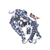


 PDBj
PDBj


 Assembly


 Mass: 583.118 Da / Num. of mol.: 4 / Source method: obtained synthetically / Formula: C29H27ClN2O5S2
 Mass: 18.015 Da / Num. of mol.: 360 / Source method: isolated from a natural source / Formula: H2O
 Sample preparation
 / Beamline: 8.3.1 / Wavelength: 1.1159 Å
 Processing