 Movie
Movie Controller

[English] 日本語
 Yorodumi
- PDB-2omd: Crystal structure of molybdopterin converting factor subunit 2 (a... -
+ Open data

- Basic information
| Entry | Database: PDB / ID: 2omd | ||||||
|---|---|---|---|---|---|---|---|
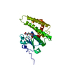
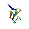
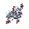
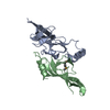
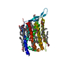
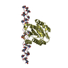
| Title | Crystal structure of molybdopterin converting factor subunit 2 (aq_2181) from aquifex aeolicus VF5 | ||||||
 Components Components | Molybdopterin-converting factor subunit 2 | ||||||
 Keywords Keywords | LYASE / MOAE / coenzyme biosynthesis / Structural Genomics / NPPSFA / National Project on Protein Structural and Functional Analyses / RIKEN Structural Genomics/Proteomics Initiative / RSGI | ||||||
| Function / homology |  Function and homology information Function and homology informationmolybdopterin synthase activity / molybdopterin synthase / Mo-molybdopterin cofactor biosynthetic process / cytosol Similarity search - Function | ||||||
| Biological species |   Aquifex aeolicus (bacteria) Aquifex aeolicus (bacteria) | ||||||
| Method |  X-RAY DIFFRACTION / SYNCHROTRON / SAD / Resolution: 2 Å X-RAY DIFFRACTION / SYNCHROTRON / SAD / Resolution: 2 Å | ||||||
 Authors Authors | Jeyakanthan, J. / Kanaujia, S.P. / Vasuki Ranjani, C. / Sekar, K. / Agari, Y. / Ebihara, A. / Kuramitsu, S. / Shinkai, A. / Shiro, Y. / Yokoyama, S. / RIKEN Structural Genomics/Proteomics Initiative (RSGI) | ||||||
 Citation Citation | Journal: To be Published Title: Crystal structure of molybdopterin converting factor subunit 2 (aq_2181) from aquifex aeolicus VF5 Authors: Jeyakanthan, J. / Kanaujia, S.P. / Vasuki Ranjani, C. / Sekar, K. / Agari, Y. / Ebihara, A. / Kuramitsu, S. / Shinkai, A. / Shiro, Y. / Yokoyama, S. | ||||||
| History |
|
- Structure visualization
| Structure viewer | Molecule: MolmilJmol/JSmol |
|---|
- Downloads & links
-Download
| PDBx/mmCIF format | 2omd.cif.gz | 79.4 KB | Display | PDBx/mmCIF format |
|---|---|---|---|---|
| PDB format | pdb2omd.ent.gz | 59.4 KB | Display | PDB format |
| PDBx/mmJSON format | 2omd.json.gz | Tree view | PDBx/mmJSON format | |
| Others |  Other downloads Other downloads |
-Validation report
| Arichive directory | https://data.pdbj.org/pub/pdb/validation_reports/om/2omdftp://data.pdbj.org/pub/pdb/validation_reports/om/2omd | HTTPS FTP |
|---|
-Related structure data
| Similar structure data | |
|---|---|
| Other databases |


-Links
 PDBj
PDBj- Assembly
| Deposited unit | 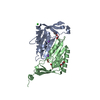
| ||||||||
|---|---|---|---|---|---|---|---|---|---|
| 1 |
| ||||||||
| Unit cell |
|
-Components
-Protein , 1 types, 2 molecules AB
| #1: Protein | Mass: 17606.289 Da / Num. of mol.: 2 Source method: isolated from a genetically manipulated source Source: (gene. exp.) Aquifex aeolicus (bacteria) / Strain: VF5 / Plasmid: PET21d / Production host: |
|---|
-Non-polymers , 6 types, 387 molecules 


| #2: Chemical |  Mass: 22.990 Da / Num. of mol.: 2 / Source method: obtained synthetically / Formula: Na Mass: 22.990 Da / Num. of mol.: 2 / Source method: obtained synthetically / Formula: Na#3: Chemical | ChemComp-CL / |  Mass: 35.453 Da / Num. of mol.: 1 / Source method: obtained synthetically / Formula: Cl Mass: 35.453 Da / Num. of mol.: 1 / Source method: obtained synthetically / Formula: Cl#4: Chemical | ChemComp-TRS / |  Mass: 122.143 Da / Num. of mol.: 1 / Source method: obtained synthetically / Formula: C4H12NO3 / Comment: pH buffer*YM Mass: 122.143 Da / Num. of mol.: 1 / Source method: obtained synthetically / Formula: C4H12NO3 / Comment: pH buffer*YM#5: Chemical |  Mass: 46.025 Da / Num. of mol.: 2 / Source method: obtained synthetically / Formula: CH2O2 Mass: 46.025 Da / Num. of mol.: 2 / Source method: obtained synthetically / Formula: CH2O2#6: Chemical |  Mass: 92.094 Da / Num. of mol.: 2 / Source method: obtained synthetically / Formula: C3H8O3 Mass: 92.094 Da / Num. of mol.: 2 / Source method: obtained synthetically / Formula: C3H8O3#7: Water | ChemComp-HOH / | Mass: 18.015 Da / Num. of mol.: 379 / Source method: isolated from a natural source / Formula: H2O |
|---|
-Experimental details
-Experiment
| Experiment | Method: X-RAY DIFFRACTION / Number of used crystals: 1 |
|---|
- Sample preparation
| Crystal | Density Matthews: 3.71 Å3/Da / Density % sol: 66.86 % |
|---|---|
| Crystal grow | Temperature: 291 K / Method: microbatch Details: 10% 1,2-propanediol, 25% Glycerol, MICROBATCH, temperature 291K |
-Data collection
| Diffraction | Mean temperature: 100 K | ||||||||||||
|---|---|---|---|---|---|---|---|---|---|---|---|---|---|
| Diffraction source | Source: SYNCHROTRON / Site: SPring-8  / Beamline: BL26B2 / Wavelength: 0.9788, 0.9000, 0.9794 / Beamline: BL26B2 / Wavelength: 0.9788, 0.9000, 0.9794 | ||||||||||||
| Detector | Type: RIGAKU RAXIS V / Detector: IMAGE PLATE / Date: Oct 15, 2006 / Details: RH Coated Bent-Cyrindrical MIRROR | ||||||||||||
| Radiation | Monochromator: SI 1 1 1 Double Crystal Monochromator / Protocol: MAD / Monochromatic (M) / Laue (L): M / Scattering type: x-ray | ||||||||||||
| Radiation wavelength |
| ||||||||||||
| Reflection | Resolution: 2→50 Å / Num. obs: 36659 / % possible obs: 100 % / Biso Wilson estimate: 25.6 Å2 / Rmerge(I) obs: 0.067 / Rsym value: 0.073 | ||||||||||||
| Reflection shell | Resolution: 2→2.07 Å / Rmerge(I) obs: 0.249 / Num. unique all: 3583 / Rsym value: 0.264 / % possible all: 100 |
- Processing
| Software |
| ||||||||||||||||||||
|---|---|---|---|---|---|---|---|---|---|---|---|---|---|---|---|---|---|---|---|---|---|
| Refinement | Method to determine structure: SAD / Resolution: 2→38.89 Å / Rfactor Rfree error: 0.004 / Data cutoff high absF: 1319976.37 / Data cutoff low absF: 0 / Isotropic thermal model: RESTRAINED / Cross valid method: THROUGHOUT / σ(F): 0 / Stereochemistry target values: Engh & Huber
| ||||||||||||||||||||
| Solvent computation | Solvent model: FLAT MODEL / Bsol: 59.8037 Å2 / ksol: 0.339765 e/Å3 | ||||||||||||||||||||
| Displacement parameters | Biso mean: 32 Å2
| ||||||||||||||||||||
| Refine analyze |
| ||||||||||||||||||||
| Refinement step | Cycle: LAST / Resolution: 2→38.89 Å
| ||||||||||||||||||||
| Refine LS restraints |
| ||||||||||||||||||||
| LS refinement shell | Resolution: 2→2.09 Å / Rfactor Rfree error: 0.012 / Total num. of bins used: 8
| ||||||||||||||||||||
| Xplor file |
|
