 Movie
Movie Controller

[English] 日本語
 Yorodumi
- PDB-2oey: Solution Structure of a Designed Spirocyclic Helical Ligand Bindi... -
+ Open data

- Basic information
| Entry | Database: PDB / ID: 2oey | ||||||
|---|---|---|---|---|---|---|---|
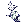
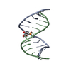
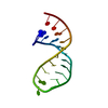
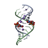
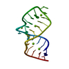
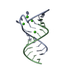
| Title | Solution Structure of a Designed Spirocyclic Helical Ligand Binding at a Two-Base Bulge Site in DNA | ||||||
 Components Components | DNA (25-MER) | ||||||
 Keywords Keywords | DNA / Designed spirocyclic helical ligand-bulged DNA complex | ||||||
| Function / homology | Chem-S2A / DNA / DNA (> 10) Function and homology information Function and homology information | ||||||
| Method | SOLUTION NMR / simulated annealing, molecular dynamics | ||||||
 Authors Authors | Zhang, N. / Lin, Y. / Xiao, Z. / Jones, G.B. / Goldberg, I.H. | ||||||
 Citation Citation | Journal: Biochemistry / Year: 2007 Title: Solution Structure of a Designed Spirocyclic Helical Ligand Binding at a Two-Base Bulge Site in DNA. Authors: Zhang, N. / Lin, Y. / Xiao, Z. / Jones, G.B. / Goldberg, I.H. #1: Journal: Science / Year: 1996Title: Solution structure of a two-base DNA bulge complexed with an enediyne cleaving analogue Authors: Stassinopoulos, A. / Ji, J. / Gao, X. / Goldberg, I.H. #2: Journal: Biochemistry / Year: 2002Title: Induced formation of a DNA bulge structure by a molecular wedge ligand-post activated neocarzinostatin chromophore Authors: Gao, X. / Stassinopolous, A. / Ji, J. / Kwon, Y. / Bare, S. / Goldberg, I.H. | ||||||
| History |
|
- Structure visualization
| Structure viewer | Molecule: MolmilJmol/JSmol |
|---|
- Downloads & links
-Download
| PDBx/mmCIF format | 2oey.cif.gz | 150 KB | Display | PDBx/mmCIF format |
|---|---|---|---|---|
| PDB format | pdb2oey.ent.gz | 118.3 KB | Display | PDB format |
| PDBx/mmJSON format | 2oey.json.gz | Tree view | PDBx/mmJSON format | |
| Others |  Other downloads Other downloads |
-Validation report
| Arichive directory | https://data.pdbj.org/pub/pdb/validation_reports/oe/2oeyftp://data.pdbj.org/pub/pdb/validation_reports/oe/2oey | HTTPS FTP |
|---|
-Related structure data
| Similar structure data |
|---|
-Links
 PDBj
PDBj


- Assembly
| Deposited unit | 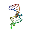
| |||||||||
|---|---|---|---|---|---|---|---|---|---|---|
| 1 |
| |||||||||
| NMR ensembles |
|
-Components
| #1: DNA chain | Mass: 7680.945 Da / Num. of mol.: 1 / Source method: obtained synthetically / Details: This is an artifical sequence |
|---|---|
| #2: Chemical | ChemComp-S2A / (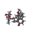  Mass: 539.618 Da / Num. of mol.: 1 / Source method: obtained synthetically / Formula: C33H33NO6 Mass: 539.618 Da / Num. of mol.: 1 / Source method: obtained synthetically / Formula: C33H33NO6 |
-Experimental details
-Experiment
| Experiment | Method: SOLUTION NMR | ||||||||||||||||||||
|---|---|---|---|---|---|---|---|---|---|---|---|---|---|---|---|---|---|---|---|---|---|
| NMR experiment |
| ||||||||||||||||||||
| NMR details | Text: This structure was determined using standard methods for DNA-drug complex. |
- Sample preparation
| Details |
| |||||||||||||||||||||||||
|---|---|---|---|---|---|---|---|---|---|---|---|---|---|---|---|---|---|---|---|---|---|---|---|---|---|---|
| Sample conditions |
|
-NMR measurement
| Radiation | Protocol: SINGLE WAVELENGTH / Monochromatic (M) / Laue (L): M |
|---|---|
| Radiation wavelength | Relative weight: 1 |
| NMR spectrometer | Type: Varian INOVA / Manufacturer: Varian / Model: INOVA / Field strength: 600 MHz |
- Processing
| NMR software |
| ||||||||||||
|---|---|---|---|---|---|---|---|---|---|---|---|---|---|
| Refinement | Method: simulated annealing, molecular dynamics / Software ordinal: 1 Details: the structures are based on a total of 531 restraints, 506 are NOE-derived distance constraints, 25 dihedral angle restraints,40 distance restraints from hydrogen bonds. | ||||||||||||
| NMR representative | Selection criteria: lowest energy | ||||||||||||
| NMR ensemble | Conformer selection criteria: back calculated data agree with experimental NOESY spectrum Conformers calculated total number: 10 / Conformers submitted total number: 8 |
 X-PLOR