 Movie
Movie Controller

[English] 日本語
 Yorodumi
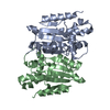
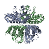
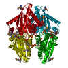
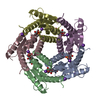
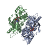
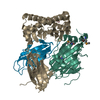
- PDB-2o6h: Lumazine synthase RibH1 from Brucella melitensis (Gene BMEI1187, ... -
+ Open data

- Basic information
| Entry | Database: PDB / ID: 2o6h | ||||||
|---|---|---|---|---|---|---|---|
| Title | Lumazine synthase RibH1 from Brucella melitensis (Gene BMEI1187, Swiss-Prot entry Q8YGH2) complexed with inhibitor 5-Nitro-6-(D-Ribitylamino)-2,4(1H,3H) Pyrimidinedione | ||||||
 Components Components | 6,7-dimethyl-8-ribityllumazine synthase 1 | ||||||
 Keywords Keywords | TRANSFERASE / lumazine synthase | ||||||
| Function / homology |  Function and homology information Function and homology information6,7-dimethyl-8-ribityllumazine synthase / 6,7-dimethyl-8-ribityllumazine synthase activity / riboflavin synthase complex / riboflavin biosynthetic process / cytosol Similarity search - Function | ||||||
| Biological species |  Brucella melitensis (bacteria) Brucella melitensis (bacteria) | ||||||
| Method |  X-RAY DIFFRACTION / SYNCHROTRON / MOLECULAR REPLACEMENT / Resolution: 2.7 Å X-RAY DIFFRACTION / SYNCHROTRON / MOLECULAR REPLACEMENT / Resolution: 2.7 Å | ||||||
 Authors Authors | Klinke, S. / Zylberman, V. / Bonomi, H.R. / Haase, I. / Guimaraes, B.G. / Braden, B.C. / Bacher, A. / Fischer, M. / Goldbaum, F.A. | ||||||
 Citation Citation | Journal: J.Mol.Biol. / Year: 2007 Title: Structural and Kinetic Properties of Lumazine Synthase Isoenzymes in the Order Rhizobiales Authors: Klinke, S. / Zylberman, V. / Bonomi, H.R. / Haase, I. / Guimaraes, B.G. / Braden, B.C. / Bacher, A. / Fischer, M. / Goldbaum, F.A. | ||||||
| History |
|
- Structure visualization
| Structure viewer | Molecule: MolmilJmol/JSmol |
|---|
- Downloads & links
-Download
| PDBx/mmCIF format | 2o6h.cif.gz | 149 KB | Display | PDBx/mmCIF format |
|---|---|---|---|---|
| PDB format | pdb2o6h.ent.gz | 118.6 KB | Display | PDB format |
| PDBx/mmJSON format | 2o6h.json.gz | Tree view | PDBx/mmJSON format | |
| Others |  Other downloads Other downloads |
-Validation report
| Arichive directory | https://data.pdbj.org/pub/pdb/validation_reports/o6/2o6hftp://data.pdbj.org/pub/pdb/validation_reports/o6/2o6h | HTTPS FTP |
|---|
-Related structure data
| Related structure data | 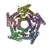 2f59SC 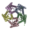 2i0fC 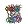 2obxC S: Starting model for refinement C: citing same article ( |
|---|---|
| Similar structure data |


-Links
 PDBj
PDBj- Assembly
| Deposited unit | 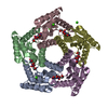
| ||||||||
|---|---|---|---|---|---|---|---|---|---|
| 1 |
| ||||||||
| 2 | 
| ||||||||
| Unit cell |
|
-Components
| #1: Protein | Mass: 16826.791 Da / Num. of mol.: 5 Source method: isolated from a genetically manipulated source Source: (gene. exp.) Brucella melitensis (bacteria) / Gene: ribH1 / Plasmid: pET11A / Species (production host): Escherichia coli / Production host: References: UniProt: Q8YGH2, 6,7-dimethyl-8-ribityllumazine synthase #2: Chemical | ChemComp-CA /   Mass: 40.078 Da / Num. of mol.: 6 / Source method: obtained synthetically / Formula: Ca Mass: 40.078 Da / Num. of mol.: 6 / Source method: obtained synthetically / Formula: Ca#3: Chemical | ChemComp-INI /   Mass: 306.229 Da / Num. of mol.: 5 / Source method: obtained synthetically / Formula: C9H14N4O8 Mass: 306.229 Da / Num. of mol.: 5 / Source method: obtained synthetically / Formula: C9H14N4O8#4: Water | ChemComp-HOH / |  Mass: 18.015 Da / Num. of mol.: 87 / Source method: isolated from a natural source / Formula: H2O Mass: 18.015 Da / Num. of mol.: 87 / Source method: isolated from a natural source / Formula: H2O |
|---|
-Experimental details
-Experiment
| Experiment | Method: X-RAY DIFFRACTION / Number of used crystals: 1 |
|---|
- Sample preparation
| Crystal | Density Matthews: 2.87 Å3/Da / Density % sol: 57.12 % |
|---|---|
| Crystal grow | Temperature: 292 K / Method: vapor diffusion, hanging drop / pH: 6.3 Details: 22% PEG 400, 0.2M CaCl2, 0.1M MES, pH 6.3, VAPOR DIFFUSION, HANGING DROP, temperature 292K |
-Data collection
| Diffraction | Mean temperature: 100 K |
|---|---|
| Diffraction source | Source: SYNCHROTRON / Site: NSLS  / Beamline: X9A / Wavelength: 0.9794 Å / Beamline: X9A / Wavelength: 0.9794 Å |
| Detector | Type: MAR CCD 165 mm / Detector: CCD / Date: Apr 26, 2006 |
| Radiation | Monochromator: Si double crystal monocromator / Protocol: SINGLE WAVELENGTH / Monochromatic (M) / Laue (L): M / Scattering type: x-ray |
| Radiation wavelength | Wavelength: 0.9794 Å / Relative weight: 1 |
| Reflection | Resolution: 2.7→48.8 Å / Num. all: 27278 / Num. obs: 22799 / % possible obs: 83.5 % / Redundancy: 3.9 % / Biso Wilson estimate: 54.6 Å2 / Rmerge(I) obs: 0.1 / Rsym value: 0.1 / Net I/σ(I): 5.8 |
| Reflection shell | Resolution: 2.7→2.85 Å / Redundancy: 2.2 % / Rmerge(I) obs: 0.313 / Mean I/σ(I) obs: 2.2 / Num. unique all: 3174 / Rsym value: 0.313 / % possible all: 82.3 |
- Processing
| Software |
| |||||||||||||||||||||||||
|---|---|---|---|---|---|---|---|---|---|---|---|---|---|---|---|---|---|---|---|---|---|---|---|---|---|---|
| Refinement | Method to determine structure: MOLECULAR REPLACEMENT Starting model: PDB ENTRY 2F59 Resolution: 2.7→48.8 Å / Cross valid method: THROUGHOUT / σ(F): 2 / Stereochemistry target values: Engh & Huber
| |||||||||||||||||||||||||
| Displacement parameters | Biso mean: 32.68 Å2 | |||||||||||||||||||||||||
| Refinement step | Cycle: LAST / Resolution: 2.7→48.8 Å
| |||||||||||||||||||||||||
| Refine LS restraints |
|
