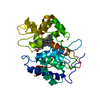
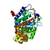
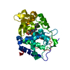
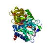
BIOMOLECULE: 1,2 THIS ENTRY CONTAINS THE CRYSTALLOGRAPHIC ASYMMETRIC UNIT WHICH CONSISTS OF 2 ...BIOMOLECULE: 1,2 THIS ENTRY CONTAINS THE CRYSTALLOGRAPHIC ASYMMETRIC UNIT WHICH CONSISTS OF 2 CHAIN(S). SEE REMARK 350 FOR INFORMATION ON GENERATING THE BIOLOGICAL MOLECULE(S). SIZE EXCLUSION CHROMATOGRAPHY WITH STATIC LIGHT SCATTERING SUPPORTS THE ASSIGNMENT OF A MONOMER AS A BIOLOGICALLY SIGNIFICANT OLIGOMERIZATION STATE.
Remark 999
SEQUENCE (1) THE CONSTRUCT WAS EXPRESSED WITH A PURIFICATION TAG MGSDKIHHHHHHENLYFQG. THE TAG WAS ...SEQUENCE (1) THE CONSTRUCT WAS EXPRESSED WITH A PURIFICATION TAG MGSDKIHHHHHHENLYFQG. THE TAG WAS REMOVED WITH TEV PROTEASE LEAVING ONLY A GLYCINE, FOLLOWED BY THE TARGET SEQUENCE. (2) THE SEQUENCE OF THE PROTEIN WAS NOT AVAILABLE AT UNP DATABASE AT THE TIME OF PROCESSING. (3) THE SEQUENCE IS AVAILABLE FROM GENBANK UNDER ACCESSION ID ZP_00105914.2 AND FROM THE UNIPROT ARCHIVE UNDER ACCESSION ID UPI000045C1C7.
Type: MARMOSAIC 325 mm CCD / Detector: CCD / Date: Nov 20, 2006 / Details: Flat mirror (vertical focusing)
Radiation
Monochromator: Single crystal Si(111) bent monochromator (horizontal focusing) Protocol: SINGLE WAVELENGTH / Monochromatic (M) / Laue (L): M / Scattering type: x-ray
Radiation wavelength
Wavelength: 0.97939 Å / Relative weight: 1
Reflection
Resolution: 1.75→29.148 Å / Num. obs: 56361 / % possible obs: 93.7 % / Biso Wilson estimate: 24.116 Å2 / Rmerge(I) obs: 0.083 / Net I/σ(I): 9.71
Reflection shell
Resolution (Å)
Rmerge(I) obs
Mean I/σ(I) obs
Num. measured obs
Diffraction-ID
% possible all
1.75-1.81
0.462
1.6
14596
1
70.4
1.81-1.89
0.383
2.4
23510
1
80.4
1.89-1.97
0.292
3.7
26700
1
89.3
1.97-2.07
0.244
5.2
39530
1
98.9
2.07-2.2
0.202
6.4
43832
1
99.8
2.2-2.37
0.169
7.6
43734
1
99.9
2.37-2.61
0.134
9.2
44214
1
99.9
2.61-2.99
0.097
12.4
44060
1
99.9
2.99-29.1
0.054
18.9
43890
1
99.8
-
Phasing
Phasing
Method: SAD
-
Processing
Software
Name
Version
Classification
NB
MolProbity
3beta29
modelbuilding
REFMAC
5.2.0005
refinement
XSCALE
datascaling
PDB_EXTRACT
2
dataextraction
XDS
datareduction
SHARP
phasing
SOLVE
phasing
Refinement
Method to determine structure: SAD / Resolution: 1.75→29.148 Å / Cor.coef. Fo:Fc: 0.964 / Cor.coef. Fo:Fc free: 0.942 / SU B: 5.28 / SU ML: 0.084 / TLS residual ADP flag: LIKELY RESIDUAL / Cross valid method: THROUGHOUT / σ(F): 0 / ESU R: 0.114 / ESU R Free: 0.114 Stereochemistry target values: MAXIMUM LIKELIHOOD WITH PHASES Details: 1. HYDROGENS HAVE BEEN ADDED IN THE RIDING POSITIONS. 2. ATOM RECORD CONTAINS RESIDUAL B FACTORS ONLY. 3. A MET-INHIBITION PROTOCOL WAS USED FOR SELENOMETHIONINE INCORPORATION DURING PROTEIN ...Details: 1. HYDROGENS HAVE BEEN ADDED IN THE RIDING POSITIONS. 2. ATOM RECORD CONTAINS RESIDUAL B FACTORS ONLY. 3. A MET-INHIBITION PROTOCOL WAS USED FOR SELENOMETHIONINE INCORPORATION DURING PROTEIN EXPRESSION. THE OCCUPANCY OF THE SE ATOMS IN THE MSE RESIDUES WAS REDUCED TO 0.75 FOR THE REDUCED SCATTERING POWER DUE TO PARTIAL S-MET INCORPORATION. 4. CHLORINE AND GLYCEROL MODELED BASED ON CRYSTALLIZATION CONDITIONS. 5. RESIDUES B182-186 HAVE VERY POOR DENSITY, AND WERE MODELED BASED ON THE CORRESPONDING RESDIUES IN CHAIN A.
Rfactor
Num. reflection
% reflection
Selection details
Rfree
0.208
2834
5 %
RANDOM
Rwork
0.166
-
-
-
obs
0.168
56308
93.94 %
-
Solvent computation
Ion probe radii: 0.8 Å / Shrinkage radii: 0.8 Å / VDW probe radii: 1.2 Å / Solvent model: MASK
Displacement parameters
Biso mean: 21.992 Å2
Baniso -1
Baniso -2
Baniso -3
1-
2.08 Å2
0 Å2
0 Å2
2-
-
-1.18 Å2
0 Å2
3-
-
-
-0.9 Å2
Refinement step
Cycle: LAST / Resolution: 1.75→29.148 Å
Protein
Nucleic acid
Ligand
Solvent
Total
Num. atoms
4225
0
92
506
4823
Refine LS restraints
Refine-ID
Type
Dev ideal
Dev ideal target
Number
X-RAY DIFFRACTION
r_bond_refined_d
0.016
0.022
4440
X-RAY DIFFRACTION
r_bond_other_d
0.002
0.02
4106
X-RAY DIFFRACTION
r_angle_refined_deg
1.641
1.981
6004
X-RAY DIFFRACTION
r_angle_other_deg
0.828
3
9520
X-RAY DIFFRACTION
r_dihedral_angle_1_deg
6.706
5
556
X-RAY DIFFRACTION
r_dihedral_angle_2_deg
34.783
23.832
214
X-RAY DIFFRACTION
r_dihedral_angle_3_deg
12.507
15
756
X-RAY DIFFRACTION
r_dihedral_angle_4_deg
18.369
15
41
X-RAY DIFFRACTION
r_chiral_restr
0.102
0.2
668
X-RAY DIFFRACTION
r_gen_planes_refined
0.007
0.02
4927
X-RAY DIFFRACTION
r_gen_planes_other
0.001
0.02
916
X-RAY DIFFRACTION
r_nbd_refined
0.222
0.2
626
X-RAY DIFFRACTION
r_nbd_other
0.204
0.2
4346
X-RAY DIFFRACTION
r_nbtor_refined
0.176
0.2
2072
X-RAY DIFFRACTION
r_nbtor_other
0.084
0.2
3076
X-RAY DIFFRACTION
r_xyhbond_nbd_refined
0.182
0.2
345
X-RAY DIFFRACTION
r_symmetry_vdw_refined
0.276
0.2
14
X-RAY DIFFRACTION
r_symmetry_vdw_other
0.247
0.2
88
X-RAY DIFFRACTION
r_symmetry_hbond_refined
0.158
0.2
20
X-RAY DIFFRACTION
r_mcbond_it
1.059
1.5
2783
X-RAY DIFFRACTION
r_mcbond_other
0.267
1.5
1118
X-RAY DIFFRACTION
r_mcangle_it
1.636
2
4338
X-RAY DIFFRACTION
r_scbond_it
2.645
3
1873
X-RAY DIFFRACTION
r_scangle_it
4.174
4.5
1656
LS refinement shell
Resolution: 1.748→1.794 Å / Total num. of bins used: 20
Rfactor
Num. reflection
% reflection
Rfree
0.271
164
-
Rwork
0.246
2888
-
obs
-
3052
70.34 %
Refinement TLS params.
Method: refined / Refine-ID: X-RAY DIFFRACTION
ID
L11 (°2)
L12 (°2)
L13 (°2)
L22 (°2)
L23 (°2)
L33 (°2)
S11 (Å °)
S12 (Å °)
S13 (Å °)
S21 (Å °)
S22 (Å °)
S23 (Å °)
S31 (Å °)
S32 (Å °)
S33 (Å °)
T11 (Å2)
T12 (Å2)
T13 (Å2)
T22 (Å2)
T23 (Å2)
T33 (Å2)
Origin x (Å)
Origin y (Å)
Origin z (Å)
1
0.6585
0.458
-0.2283
1.4993
-0.2022
0.5635
0.032
-0.068
-0.0058
0.0363
-0.0325
-0.167
-0.0363
0.1386
0.0005
-0.0674
0.0069
0.004
-0.0759
0.0033
-0.0947
15.543
16.631
31.559
2
0.6903
0.4863
-0.0389
1.4781
0.079
1.3507
0.001
-0.0022
-0.0571
0.0228
-0.0084
0.1605
0.0233
-0.0805
0.0073
-0.0786
0.0157
0.0015
-0.1149
0.0026
-0.0874
0.024
4.623
27.64
3
0.6658
0.1889
-0.0077
2.3142
-0.399
1.4353
0.0163
0.0237
-0.1117
-0.021
-0.0345
0.1376
0.1425
-0.1046
0.0182
-0.104
0.0052
-0.0077
-0.0549
-0.0043
-0.0723
16.784
-4.036
59.427
4
1.3429
0.353
0.5968
1.8692
0.5164
1.0104
0.0115
0.1079
0.0886
-0.1314
-0.0157
-0.1244
-0.1049
0.2255
0.0042
-0.0847
-0.0043
0.0265
-0.0236
0.0204
-0.0645
31.426
9.407
62.161
Refinement TLS group
Refine-ID: X-RAY DIFFRACTION / Selection: ALL
ID
Refine TLS-ID
Auth asym-ID
Label asym-ID
Auth seq-ID
Label seq-ID
1
1
A
A
2 - 132
3 - 133
2
2
A
A
133 - 269
134 - 270
3
3
B
B
2 - 132
3 - 133
4
4
B
B
133 - 269
134 - 270
+
About Yorodumi
-
News
-
Feb 9, 2022. New format data for meta-information of EMDB entries
New format data for meta-information of EMDB entries
Version 3 of the EMDB header file is now the official format.
The previous official version 1.9 will be removed from the archive.
In the structure databanks used in Yorodumi, some data are registered as the other names, "COVID-19 virus" and "2019-nCoV". Here are the details of the virus and the list of structure data.
Jan 31, 2019. EMDB accession codes are about to change! (news from PDBe EMDB page)
EMDB accession codes are about to change! (news from PDBe EMDB page)
The allocation of 4 digits for EMDB accession codes will soon come to an end. Whilst these codes will remain in use, new EMDB accession codes will include an additional digit and will expand incrementally as the available range of codes is exhausted. The current 4-digit format prefixed with “EMD-” (i.e. EMD-XXXX) will advance to a 5-digit format (i.e. EMD-XXXXX), and so on. It is currently estimated that the 4-digit codes will be depleted around Spring 2019, at which point the 5-digit format will come into force.
The EM Navigator/Yorodumi systems omit the EMD- prefix.
Related info.:Q: What is EMD? / ID/Accession-code notation in Yorodumi/EM Navigator
Yorodumi is a browser for structure data from EMDB, PDB, SASBDB, etc.
This page is also the successor to EM Navigator detail page, and also detail information page/front-end page for Omokage search.
The word "yorodu" (or yorozu) is an old Japanese word meaning "ten thousand". "mi" (miru) is to see.
Related info.:EMDB / PDB / SASBDB / Comparison of 3 databanks / Yorodumi Search / Aug 31, 2016. New EM Navigator & Yorodumi / Yorodumi Papers / Jmol/JSmol / Function and homology information / Changes in new EM Navigator and Yorodumi
 Movie
Movie Controller

 Yorodumi
 Open data
 Basic information
 Components
 Keywords
 Function and homology information
 Nostoc punctiforme (bacteria)
 X-RAY DIFFRACTION /
  Authors
 Citation
 Structure visualization
 Downloads & links
 Other downloads

 PDBj
PDBj

 Assembly


 Mass: 35.453 Da / Num. of mol.: 2 / Source method: obtained synthetically / Formula: Cl
 Mass: 92.094 Da / Num. of mol.: 15 / Source method: obtained synthetically / Formula: C3H8O3
 Mass: 18.015 Da / Num. of mol.: 506 / Source method: isolated from a natural source / Formula: H2O
 Sample preparation
 / Beamline: BL11-1 / Wavelength: 0.97939
  Processing