- PDB-2o4t: CRYSTAL STRUCTURE OF a protein of the DUF1048 family with a left-... -
+
Open data
ID or keywords:
Loading...
-
Basic information
Entry
Database: PDB / ID: 2o4t
Title
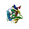
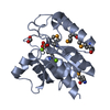
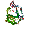
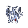
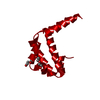
CRYSTAL STRUCTURE OF a protein of the DUF1048 family with a left-handed superhelix fold (BH3976) FROM BACILLUS HALODURANS AT 1.95 A RESOLUTION
Components
BH3976 protein
Keywords
UNKNOWN FUNCTION / LEFT-HANDED SUPERHELIX FOLD / STRUCTURAL GENOMICS / JOINT CENTER FOR STRUCTURAL GENOMICS / JCSG / PROTEIN STRUCTURE INITIATIVE / PSI-2
Function / homology
Uncharacterised conserved protein UCP029876 / Protein of unknown function (DUF1048) / c-terminal domain of poly(a) binding protein / c-terminal domain of poly(a) binding protein / Orthogonal Bundle / Mainly Alpha / DI(HYDROXYETHYL)ETHER / BH3976 protein
BIOMOLECULE: 1 THIS ENTRY CONTAINS THE CRYSTALLOGRAPHIC ASYMMETRIC UNIT WHICH CONSISTS OF 1 CHAIN(S) ...BIOMOLECULE: 1 THIS ENTRY CONTAINS THE CRYSTALLOGRAPHIC ASYMMETRIC UNIT WHICH CONSISTS OF 1 CHAIN(S). SEE REMARK 350 FOR INFORMATION ON GENERATING THE BIOLOGICAL MOLECULE(S). SIZE EXCLUSION CHROMATOGRAPHY WITH STATIC LIGHT SCATTERING SUPPORTS THE ASSIGNMENT OF A DIMER AS A BIOLOGICALLY SIGNIFICANT OLIGOMERIZATION STATE.
Remark 999
SEQUENCE THE CONSTRUCT WAS EXPRESSED WITH A PURIFICATION TAG MGSDKIHHHHHHENLYFQG. THE TAG WAS ...SEQUENCE THE CONSTRUCT WAS EXPRESSED WITH A PURIFICATION TAG MGSDKIHHHHHHENLYFQG. THE TAG WAS REMOVED WITH TEV PROTEASE LEAVING ONLY A GLYCINE (0) FOLLOWED BY RESIDUE 15 OF THE TARGET SEQUENCE.
Resolution: 1.95→60.412 Å / Cor.coef. Fo:Fc: 0.958 / Cor.coef. Fo:Fc free: 0.92 / SU B: 8.919 / SU ML: 0.124 / TLS residual ADP flag: LIKELY RESIDUAL / Cross valid method: THROUGHOUT / σ(F): 0 / ESU R: 0.131 / ESU R Free: 0.138 / Stereochemistry target values: MAXIMUM LIKELIHOOD Details: 1. HYDROGENS HAVE BEEN ADDED IN THE RIDING POSITIONS. 2. ATOM RECORD CONTAINS RESIDUAL B FACTORS ONLY. 3. ELECTRON DENSITIES FOR RESIDUE 15 AND RESIDUE 106-113 WERE DISORDERED, THEREFORE ...Details: 1. HYDROGENS HAVE BEEN ADDED IN THE RIDING POSITIONS. 2. ATOM RECORD CONTAINS RESIDUAL B FACTORS ONLY. 3. ELECTRON DENSITIES FOR RESIDUE 15 AND RESIDUE 106-113 WERE DISORDERED, THEREFORE THESE RESIDUES WERE NOT MODELED. 4. TWO MOLECULES OF POLYETHYLENE GLYCOL 300 FROM THE CRYSTALLIZATION WERE MOLDELED INTO THE STRUCTURE. ONE OF THESE PEG MOLECULES IS ON A SPECIAL POSITION BETWEEN SYMMETRY-RELATED SUBUNITS.
Rfactor
Num. reflection
% reflection
Selection details
Rfree
0.255
523
4.8 %
RANDOM
Rwork
0.203
-
-
-
all
0.205
-
-
-
obs
0.205
10898
99.75 %
-
Solvent computation
Ion probe radii: 0.8 Å / Shrinkage radii: 0.8 Å / VDW probe radii: 1.2 Å / Solvent model: BABINET MODEL WITH MASK
Displacement parameters
Biso mean: 43.239 Å2
Baniso -1
Baniso -2
Baniso -3
1-
2.84 Å2
1.42 Å2
0 Å2
2-
-
2.84 Å2
0 Å2
3-
-
-
-4.25 Å2
Refinement step
Cycle: LAST / Resolution: 1.95→60.412 Å
Protein
Nucleic acid
Ligand
Solvent
Total
Num. atoms
686
0
14
56
756
Refine LS restraints
Refine-ID
Type
Dev ideal
Dev ideal target
Number
X-RAY DIFFRACTION
r_bond_refined_d
0.016
0.022
727
X-RAY DIFFRACTION
r_bond_other_d
0.002
0.02
660
X-RAY DIFFRACTION
r_angle_refined_deg
1.442
1.979
984
X-RAY DIFFRACTION
r_angle_other_deg
0.819
3
1539
X-RAY DIFFRACTION
r_dihedral_angle_1_deg
5.75
5
95
X-RAY DIFFRACTION
r_dihedral_angle_2_deg
38.084
26.176
34
X-RAY DIFFRACTION
r_dihedral_angle_3_deg
12.884
15
117
X-RAY DIFFRACTION
r_dihedral_angle_4_deg
0.682
15
1
X-RAY DIFFRACTION
r_chiral_restr
0.094
0.2
111
X-RAY DIFFRACTION
r_gen_planes_refined
0.006
0.02
819
X-RAY DIFFRACTION
r_gen_planes_other
0.001
0.02
138
X-RAY DIFFRACTION
r_nbd_refined
0.237
0.2
181
X-RAY DIFFRACTION
r_nbd_other
0.161
0.2
620
X-RAY DIFFRACTION
r_nbtor_refined
0.193
0.2
378
X-RAY DIFFRACTION
r_nbtor_other
0.088
0.2
404
X-RAY DIFFRACTION
r_xyhbond_nbd_refined
0.199
0.2
43
X-RAY DIFFRACTION
r_xyhbond_nbd_other
0.035
0.2
1
X-RAY DIFFRACTION
r_symmetry_vdw_refined
0.284
0.2
24
X-RAY DIFFRACTION
r_symmetry_vdw_other
0.173
0.2
54
X-RAY DIFFRACTION
r_symmetry_hbond_refined
0.164
0.2
3
X-RAY DIFFRACTION
r_mcbond_it
2.502
3
495
X-RAY DIFFRACTION
r_mcbond_other
0.665
3
192
X-RAY DIFFRACTION
r_mcangle_it
3.278
5
724
X-RAY DIFFRACTION
r_scbond_it
5.26
8
301
X-RAY DIFFRACTION
r_scangle_it
6.712
11
257
LS refinement shell
Resolution: 1.95→2.001 Å / Total num. of bins used: 20
Rfactor
Num. reflection
% reflection
Rfree
0.335
33
-
Rwork
0.292
724
-
obs
-
757
98.83 %
Refinement TLS params.
Method: refined / Origin x: 18.9019 Å / Origin y: -2.1762 Å / Origin z: 47.5245 Å
11
12
13
21
22
23
31
32
33
T
-0.1647 Å2
0.0732 Å2
-0.0352 Å2
-
-0.2797 Å2
0.0015 Å2
-
-
-0.1402 Å2
L
4.5907 °2
0.902 °2
-1.4456 °2
-
2.5849 °2
0.7994 °2
-
-
5.2463 °2
S
0.179 Å °
0.0165 Å °
-0.1646 Å °
0.0956 Å °
-0.1901 Å °
0.1653 Å °
-0.2279 Å °
-0.0438 Å °
0.0111 Å °
Refinement TLS group
Selection: ALL
+
About Yorodumi
-
News
-
Feb 9, 2022. New format data for meta-information of EMDB entries
New format data for meta-information of EMDB entries
Version 3 of the EMDB header file is now the official format.
The previous official version 1.9 will be removed from the archive.
In the structure databanks used in Yorodumi, some data are registered as the other names, "COVID-19 virus" and "2019-nCoV". Here are the details of the virus and the list of structure data.
Jan 31, 2019. EMDB accession codes are about to change! (news from PDBe EMDB page)
EMDB accession codes are about to change! (news from PDBe EMDB page)
The allocation of 4 digits for EMDB accession codes will soon come to an end. Whilst these codes will remain in use, new EMDB accession codes will include an additional digit and will expand incrementally as the available range of codes is exhausted. The current 4-digit format prefixed with “EMD-” (i.e. EMD-XXXX) will advance to a 5-digit format (i.e. EMD-XXXXX), and so on. It is currently estimated that the 4-digit codes will be depleted around Spring 2019, at which point the 5-digit format will come into force.
The EM Navigator/Yorodumi systems omit the EMD- prefix.
Related info.:Q: What is EMD? / ID/Accession-code notation in Yorodumi/EM Navigator
Yorodumi is a browser for structure data from EMDB, PDB, SASBDB, etc.
This page is also the successor to EM Navigator detail page, and also detail information page/front-end page for Omokage search.
The word "yorodu" (or yorozu) is an old Japanese word meaning "ten thousand". "mi" (miru) is to see.
Related info.:EMDB / PDB / SASBDB / Comparison of 3 databanks / Yorodumi Search / Aug 31, 2016. New EM Navigator & Yorodumi / Yorodumi Papers / Jmol/JSmol / Function and homology information / Changes in new EM Navigator and Yorodumi
 Movie
Movie Controller

 Yorodumi
 Open data
 Basic information
 Components
 Keywords
 Function and homology information
 Bacillus halodurans (bacteria)
 X-RAY DIFFRACTION /
  Authors
 Citation
 Structure visualization
 Downloads & links
 Other downloads


 PDBj
PDBj Assembly

 Mass: 106.120 Da / Num. of mol.: 2 / Source method: obtained synthetically / Formula: C4H10O3
 Mass: 18.015 Da / Num. of mol.: 56 / Source method: isolated from a natural source / Formula: H2O
 Sample preparation
 / Beamline: 23-ID-D / Wavelength: 0.97942
  Processing