 Movie
Movie Controller

[English] 日本語
 Yorodumi
- PDB-2o0q: X-ray Crystal Structure of Protein CC0527 from Caulobacter cresce... -
+ Open data

- Basic information
| Entry | Database: PDB / ID: 2o0q | ||||||
|---|---|---|---|---|---|---|---|
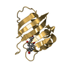
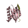
| Title | X-ray Crystal Structure of Protein CC0527 from Caulobacter crescentus. Northeast Structural Genomics Consortium Target CcR55 | ||||||
 Components Components | Hypothetical protein CC0527 | ||||||
 Keywords Keywords | STRUCTURAL GENOMICS / UNKNOWN FUNCTION / PSI / PROTEIN STRUCTURE INITIATIVE / NORTHEAST STRUCTURAL GENOMICS CONSORTIUM / NESG / Hypothetical protein CC0527 | ||||||
| Function / homology | Protein of unknown function DUF952 / Protein of unknown function DUF952 / Protein of unknown function (DUF952) / ADP-ribosylation fold / Alpha-Beta Barrel / Alpha Beta / DUF952 domain-containing protein Function and homology information Function and homology information | ||||||
| Biological species |  Caulobacter vibrioides (bacteria) Caulobacter vibrioides (bacteria) | ||||||
| Method |  X-RAY DIFFRACTION / SYNCHROTRON / MOLECULAR REPLACEMENT / Resolution: 1.8 Å X-RAY DIFFRACTION / SYNCHROTRON / MOLECULAR REPLACEMENT / Resolution: 1.8 Å | ||||||
 Authors Authors | Seetharaman, J. / Su, M. / Wang, D. / Fang, Y. / Cunningham, K. / Ma, L. / Xiao, R. / Liu, J. / Baran, M.C. / Acton, T.B. ...Seetharaman, J. / Su, M. / Wang, D. / Fang, Y. / Cunningham, K. / Ma, L. / Xiao, R. / Liu, J. / Baran, M.C. / Acton, T.B. / Rost, B. / Montelione, G.T. / Hunt, J.F. / Tong, L. / Northeast Structural Genomics Consortium (NESG) | ||||||
 Citation Citation | Journal: TO BE PUBLISHED Title: Crystal Structure of the Hypothetical Protein from Caulobacter Crescentus. Authors: Seetharaman, J. / Su, M. / Wang, D. / Fang, Y. / Cunningham, K. / Ma, L. / Xiao, R. / Liu, J. / Baran, M.C. / Acton, T.B. / Rost, B. / Montelione, G.T. / Hunt, J.F. / Tong, L. | ||||||
| History |
|
- Structure visualization
| Structure viewer | Molecule: MolmilJmol/JSmol |
|---|
- Downloads & links
-Download
| PDBx/mmCIF format | 2o0q.cif.gz | 37.5 KB | Display | PDBx/mmCIF format |
|---|---|---|---|---|
| PDB format | pdb2o0q.ent.gz | 25 KB | Display | PDB format |
| PDBx/mmJSON format | 2o0q.json.gz | Tree view | PDBx/mmJSON format | |
| Others |  Other downloads Other downloads |
-Validation report
| Arichive directory | https://data.pdbj.org/pub/pdb/validation_reports/o0/2o0qftp://data.pdbj.org/pub/pdb/validation_reports/o0/2o0q | HTTPS FTP |
|---|
-Related structure data
| Related structure data |  2o0pSC S: Starting model for refinement C: citing same article ( |
|---|---|
| Similar structure data | |
| Other databases |


-Links
 PDBj
PDBj- Assembly
| Deposited unit | 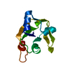
| ||||||||
|---|---|---|---|---|---|---|---|---|---|
| 1 |
| ||||||||
| Unit cell |
|
-Components
| #1: Protein | Mass: 12571.089 Da / Num. of mol.: 1 Source method: isolated from a genetically manipulated source Source: (gene. exp.) Caulobacter vibrioides (bacteria) / Production host: |
|---|---|
| #2: Water | ChemComp-HOH /  Mass: 18.015 Da / Num. of mol.: 152 / Source method: isolated from a natural source / Formula: H2O Mass: 18.015 Da / Num. of mol.: 152 / Source method: isolated from a natural source / Formula: H2O |
-Experimental details
-Experiment
| Experiment | Method: X-RAY DIFFRACTION / Number of used crystals: 1 |
|---|
- Sample preparation
| Crystal | Density Matthews: 4.07 Å3/Da / Density % sol: 69.79 % |
|---|---|
| Crystal grow | Temperature: 298 K / Method: vapor diffusion, hanging drop / pH: 5.6 Details: 0.2M Sodium acetate, 0.1M Sodium Citrate, PGE 4000 , pH 5.6, VAPOR DIFFUSION, HANGING DROP, temperature 298K |
-Data collection
| Diffraction | Mean temperature: 100 K |
|---|---|
| Diffraction source | Source: SYNCHROTRON / Site: NSLS  / Beamline: X4A / Wavelength: 0.978 Å / Beamline: X4A / Wavelength: 0.978 Å |
| Detector | Type: ADSC QUANTUM 4 / Detector: CCD / Date: Oct 10, 2006 / Details: Mirrors |
| Radiation | Monochromator: SI 111 CHANNEL / Protocol: SINGLE WAVELENGTH / Monochromatic (M) / Laue (L): M / Scattering type: x-ray |
| Radiation wavelength | Wavelength: 0.978 Å / Relative weight: 1 |
| Reflection | Resolution: 1.8→50 Å / Num. obs: 31132 / % possible obs: 98.4 % / Observed criterion σ(F): 0 / Observed criterion σ(I): 0 / Redundancy: 7.3 % / Biso Wilson estimate: 9.9 Å2 / Rmerge(I) obs: 0.07 / Rsym value: 0.054 / Net I/σ(I): 11.4 |
| Reflection shell | Resolution: 1.8→1.86 Å / Redundancy: 5.5 % / Rmerge(I) obs: 0.369 / Mean I/σ(I) obs: 9.4 / Num. unique all: 3267 / Rsym value: 0.334 / % possible all: 88.3 |
- Processing
| Software |
| ||||||||||||||||||||
|---|---|---|---|---|---|---|---|---|---|---|---|---|---|---|---|---|---|---|---|---|---|
| Refinement | Method to determine structure: MOLECULAR REPLACEMENT Starting model: 2O0P Resolution: 1.8→32.98 Å / Rfactor Rfree error: 0.007 / Data cutoff high absF: 83262.25 / Data cutoff low absF: 0 / Isotropic thermal model: RESTRAINED / Cross valid method: THROUGHOUT / σ(F): 2 / Stereochemistry target values: Engh & Huber
| ||||||||||||||||||||
| Solvent computation | Solvent model: FLAT MODEL / Bsol: 41.6208 Å2 / ksol: 0.390814 e/Å3 | ||||||||||||||||||||
| Displacement parameters | Biso mean: 21.1 Å2
| ||||||||||||||||||||
| Refine analyze |
| ||||||||||||||||||||
| Refinement step | Cycle: LAST / Resolution: 1.8→32.98 Å
| ||||||||||||||||||||
| Refine LS restraints |
| ||||||||||||||||||||
| LS refinement shell | Resolution: 1.8→1.91 Å / Rfactor Rfree error: 0.018 / Total num. of bins used: 6
| ||||||||||||||||||||
| Xplor file |
|
