 Movie
Movie Controller

[English] 日本語
 Yorodumi
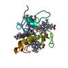
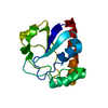
- PDB-2a8k: Structural and Mutational Studies of the Catalytic Domain of Coli... -
+ Open data

- Basic information
| Entry | Database: PDB / ID: 2a8k | ||||||
|---|---|---|---|---|---|---|---|
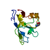
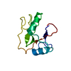
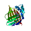
| Title | Structural and Mutational Studies of the Catalytic Domain of Colicin E5a tRNA-Specific Ribonuclease | ||||||
 Components Components | Colicin E5 | ||||||
 Keywords Keywords | HYDROLASE / Ribonuclease / Toxin | ||||||
| Function / homology |  Function and homology information Function and homology informationRNA nuclease activity / endonuclease activity / killing of cells of another organism / Hydrolases; Acting on ester bonds / defense response to bacterium / hydrolase activity Similarity search - Function | ||||||
| Biological species |  | ||||||
| Method |  X-RAY DIFFRACTION / SYNCHROTRON / MAD / Resolution: 1.5 Å X-RAY DIFFRACTION / SYNCHROTRON / MAD / Resolution: 1.5 Å | ||||||
 Authors Authors | Lin, Y.L. / Elias, Y. / Huang, R.H. | ||||||
 Citation Citation | Journal: Biochemistry / Year: 2005 Title: Structural and mutational studies of the catalytic domain of colicin E5: A tRNA-specific ribonuclease Authors: Lin, Y.L. / Elias, Y. / Huang, R.H. | ||||||
| History |
|
- Structure visualization
| Structure viewer | Molecule: MolmilJmol/JSmol |
|---|
- Downloads & links
-Download
| PDBx/mmCIF format | 2a8k.cif.gz | 92.5 KB | Display | PDBx/mmCIF format |
|---|---|---|---|---|
| PDB format | pdb2a8k.ent.gz | 70.2 KB | Display | PDB format |
| PDBx/mmJSON format | 2a8k.json.gz | Tree view | PDBx/mmJSON format | |
| Others |  Other downloads Other downloads |
-Validation report
| Arichive directory | https://data.pdbj.org/pub/pdb/validation_reports/a8/2a8kftp://data.pdbj.org/pub/pdb/validation_reports/a8/2a8k | HTTPS FTP |
|---|
-Related structure data
| Similar structure data |
|---|


-Links
 PDBj
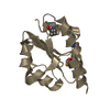
PDBj- Assembly
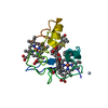
| Deposited unit | 
| ||||||||
|---|---|---|---|---|---|---|---|---|---|
| 1 | 
| ||||||||
| 2 | 
| ||||||||
| 3 | 
| ||||||||
| 4 | 
| ||||||||
| Unit cell |
|
-Components
| #1: Protein | Mass: 11996.320 Da / Num. of mol.: 4 Source method: isolated from a genetically manipulated source Source: (gene. exp.) References: UniProt: P18000, Hydrolases; Acting on ester bonds #2: Chemical | ChemComp-CA /   Mass: 40.078 Da / Num. of mol.: 4 / Source method: obtained synthetically / Formula: Ca Mass: 40.078 Da / Num. of mol.: 4 / Source method: obtained synthetically / Formula: Ca#3: Water | ChemComp-HOH / |  Mass: 18.015 Da / Num. of mol.: 346 / Source method: isolated from a natural source / Formula: H2O Mass: 18.015 Da / Num. of mol.: 346 / Source method: isolated from a natural source / Formula: H2O |
|---|
-Experimental details
-Experiment
| Experiment | Method: X-RAY DIFFRACTION / Number of used crystals: 1 |
|---|
- Sample preparation
| Crystal | Density Matthews: 2.58 Å3/Da / Density % sol: 52.29 % |
|---|---|
| Crystal grow | Temperature: 278 K / Method: vapor diffusion, hanging drop / pH: 6.5 Details: PEG400, pH 6.5, VAPOR DIFFUSION, HANGING DROP, temperature 278K |
-Data collection
| Diffraction source | Source: SYNCHROTRON / Site: APS  / Beamline: 14-BM-C / Wavelength: 0.9794, 0.9792, 0.9686 / Beamline: 14-BM-C / Wavelength: 0.9794, 0.9792, 0.9686 | ||||||||||||
|---|---|---|---|---|---|---|---|---|---|---|---|---|---|
| Detector | Type: ADSC QUANTUM 4 / Detector: CCD / Date: Mar 7, 2003 | ||||||||||||
| Radiation | Monochromator: Yale Mirrors / Protocol: MAD / Monochromatic (M) / Laue (L): M / Scattering type: x-ray | ||||||||||||
| Radiation wavelength |
| ||||||||||||
| Reflection | Resolution: 1.5→30 Å / Num. all: 76723 / Num. obs: 73778 / % possible obs: 96.2 % / Observed criterion σ(F): 2 / Observed criterion σ(I): 2 / Redundancy: 7.4 % / Rmerge(I) obs: 0.035 / Net I/σ(I): 21.3 |
- Processing
| Software |
| ||||||||||||||||||||
|---|---|---|---|---|---|---|---|---|---|---|---|---|---|---|---|---|---|---|---|---|---|
| Refinement | Method to determine structure: MAD / Resolution: 1.5→30 Å / σ(F): 2 / Stereochemistry target values: Engh & Huber
| ||||||||||||||||||||
| Refinement step | Cycle: LAST / Resolution: 1.5→30 Å
| ||||||||||||||||||||
| Refine LS restraints |
|
