 Movie
Movie Controller


+ Open data

- Basic information
| Entry | Database: PDB / ID: 2nu8 | ||||||
|---|---|---|---|---|---|---|---|
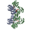
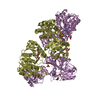
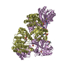
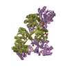
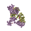
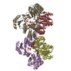
| Title | C123aT Mutant of E. coli Succinyl-CoA Synthetase | ||||||
 Components Components | (Succinyl-CoA ...) x 2 | ||||||
 Keywords Keywords | LIGASE / citric acid cycle / heterotetramer / ATP-GRASP fold / Rossmann fold | ||||||
| Function / homology |  Function and homology information Function and homology informationsuccinate-CoA ligase (GDP-forming) activity / succinate-CoA ligase complex (ADP-forming) / succinate-CoA ligase (ADP-forming) / succinate-CoA ligase complex / succinate-CoA ligase (ADP-forming) activity / succinyl-CoA metabolic process / tricarboxylic acid cycle / nucleotide binding / magnesium ion binding / ATP binding ...succinate-CoA ligase (GDP-forming) activity / succinate-CoA ligase complex (ADP-forming) / succinate-CoA ligase (ADP-forming) / succinate-CoA ligase complex / succinate-CoA ligase (ADP-forming) activity / succinyl-CoA metabolic process / tricarboxylic acid cycle / nucleotide binding / magnesium ion binding / ATP binding / cytosol / cytoplasm Similarity search - Function | ||||||
| Biological species |  | ||||||
| Method |  X-RAY DIFFRACTION / SYNCHROTRON / FOURIER SYNTHESIS / Resolution: 2.15 Å X-RAY DIFFRACTION / SYNCHROTRON / FOURIER SYNTHESIS / Resolution: 2.15 Å | ||||||
 Authors Authors | Fraser, M.E. | ||||||
 Citation Citation | Journal: ACTA CRYSTALLOGR.,SECT.D / Year: 2007 Title: Participation of Cys 123alpha of Escherichia coli Succinyl-CoA Synthetase in Catalysis Authors: Hidber, E. / Brownie, E.R. / Hayakawa, K. / Fraser, M.E. | ||||||
| History |
| ||||||
| Remark 600 | HETEROGEN Atoms missing from COA 1325 were not modeled due to lack of electron density. |
- Structure visualization
| Structure viewer | Molecule: MolmilJmol/JSmol |
|---|
- Downloads & links
-Download
| PDBx/mmCIF format | 2nu8.cif.gz | 270.7 KB | Display | PDBx/mmCIF format |
|---|---|---|---|---|
| PDB format | pdb2nu8.ent.gz | 216.4 KB | Display | PDB format |
| PDBx/mmJSON format | 2nu8.json.gz | Tree view | PDBx/mmJSON format | |
| Others |  Other downloads Other downloads |
-Validation report
| Arichive directory | https://data.pdbj.org/pub/pdb/validation_reports/nu/2nu8ftp://data.pdbj.org/pub/pdb/validation_reports/nu/2nu8 | HTTPS FTP |
|---|
-Related structure data
| Related structure data |  2nu6C  2nu7C 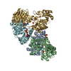 2nu9C  2nuaC 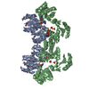 1jkjS S: Starting model for refinement C: citing same article ( |
|---|---|
| Similar structure data |


-Links
 PDBj
PDBj


- Assembly
| Deposited unit | 
| ||||||||
|---|---|---|---|---|---|---|---|---|---|
| 1 | 
| ||||||||
| 2 | 
| ||||||||
| Unit cell |
|
-Components
-Succinyl-CoA ... , 2 types, 4 molecules ADBE
| #1: Protein | Mass: 29677.201 Da / Num. of mol.: 2 / Mutation: C123T Source method: isolated from a genetically manipulated source Source: (gene. exp.) References: UniProt: P0AGE9, succinate-CoA ligase (ADP-forming) #2: Protein | Mass: 41438.496 Da / Num. of mol.: 2 Source method: isolated from a genetically manipulated source Source: (gene. exp.) References: UniProt: P0A836, succinate-CoA ligase (ADP-forming) |
|---|
-Non-polymers , 5 types, 521 molecules 


| #3: Chemical |  Mass: 94.971 Da / Num. of mol.: 2 / Source method: obtained synthetically / Formula: PO4 Mass: 94.971 Da / Num. of mol.: 2 / Source method: obtained synthetically / Formula: PO4#4: Chemical | ChemComp-SO4 /  Mass: 96.063 Da / Num. of mol.: 4 / Source method: obtained synthetically / Formula: SO4 Mass: 96.063 Da / Num. of mol.: 4 / Source method: obtained synthetically / Formula: SO4#5: Chemical | ChemComp-COA /  Mass: 767.534 Da / Num. of mol.: 4 / Source method: obtained synthetically / Formula: C21H36N7O16P3S Mass: 767.534 Da / Num. of mol.: 4 / Source method: obtained synthetically / Formula: C21H36N7O16P3S#6: Chemical | ChemComp-GOL / |  Mass: 92.094 Da / Num. of mol.: 1 / Source method: obtained synthetically / Formula: C3H8O3 Mass: 92.094 Da / Num. of mol.: 1 / Source method: obtained synthetically / Formula: C3H8O3#7: Water | ChemComp-HOH / | Mass: 18.015 Da / Num. of mol.: 510 / Source method: isolated from a natural source / Formula: H2O |
|---|
-Details
| Has protein modification | Y |
|---|
-Experimental details
-Experiment
| Experiment | Method: X-RAY DIFFRACTION / Number of used crystals: 1 |
|---|
- Sample preparation
| Crystal | Density Matthews: 3.17 Å3/Da / Density % sol: 61.19 % |
|---|---|
| Crystal grow | Temperature: 294 K / Method: vapor diffusion, hanging drop / pH: 7.9 Details: BICINE, Ammonium sulfate, pH 7.9, VAPOR DIFFUSION, HANGING DROP, temperature 294K |
-Data collection
| Diffraction | Mean temperature: 100 K |
|---|---|
| Diffraction source | Source: SYNCHROTRON / Site: CHESS  / Beamline: A1 / Wavelength: 0.9474 / Beamline: A1 / Wavelength: 0.9474 |
| Detector | Type: ADSC QUANTUM 4 / Detector: CCD / Date: Mar 4, 2002 / Details: monochromator |
| Radiation | Monochromator: Si(111) / Protocol: SINGLE WAVELENGTH / Monochromatic (M) / Laue (L): M / Scattering type: x-ray |
| Radiation wavelength | Wavelength: 0.9474 Å / Relative weight: 1 |
| Reflection | Resolution: 2.15→100 Å / Num. all: 94811 / Num. obs: 94811 / % possible obs: 89.9 % / Observed criterion σ(F): 0 / Observed criterion σ(I): 0 / Redundancy: 3.2 % / Biso Wilson estimate: 31.97 Å2 / Rmerge(I) obs: 0.14 / Net I/σ(I): 7.7 |
| Reflection shell | Resolution: 2.15→2.19 Å / Redundancy: 1.4 % / Rmerge(I) obs: 0.186 / Mean I/σ(I) obs: 1.2 / Num. unique all: 2681 / % possible all: 51.7 |
- Processing
| Software |
| |||||||||||||||||||||||||
|---|---|---|---|---|---|---|---|---|---|---|---|---|---|---|---|---|---|---|---|---|---|---|---|---|---|---|
| Refinement | Method to determine structure: FOURIER SYNTHESIS Starting model: pdb entry 1JKJ Resolution: 2.15→41.97 Å / Isotropic thermal model: Isotropic / Cross valid method: THROUGHOUT / σ(F): 0 / σ(I): 0 / Stereochemistry target values: Engh & Huber
| |||||||||||||||||||||||||
| Displacement parameters | Biso mean: 35.57 Å2
| |||||||||||||||||||||||||
| Refine analyze |
| |||||||||||||||||||||||||
| Refinement step | Cycle: LAST / Resolution: 2.15→41.97 Å
| |||||||||||||||||||||||||
| Refine LS restraints |
| |||||||||||||||||||||||||
| LS refinement shell | Resolution: 2.15→2.25 Å / Rfactor Rfree error: 0.011
|
