Mass: 18.015 Da / Num. of mol.: 10 / Source method: isolated from a natural source / Formula: H2O
Has protein modification
Y
-
Experimental details
-
Experiment
Experiment
Method: X-RAY DIFFRACTION / Number of used crystals: 1
-
Sample preparation
Crystal
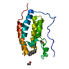
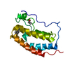
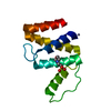
Density Matthews: 2.27 Å3/Da / Density % sol: 45.89 % Description: THE SEQUENCE DIFFERS FROM NOTEXIN'S AT 7 POSITIONS
Crystal grow
pH: 8.5 Details: THE PROTEIN WAS DISSOLVED IN 50 MM TRIS-NSULFATE, 70MM NH4AC PH 8.5; TO A TOTAL CONCENTRATION OF 15MG/ML; THE PUT IN A THICK-WALLED CAPILLARY SEALED WITH A SEMI-PERMEABLE TUBING AND PLACED ...Details: THE PROTEIN WAS DISSOLVED IN 50 MM TRIS-NSULFATE, 70MM NH4AC PH 8.5; TO A TOTAL CONCENTRATION OF 15MG/ML; THE PUT IN A THICK-WALLED CAPILLARY SEALED WITH A SEMI-PERMEABLE TUBING AND PLACED IN A VESSEL CONTAINING 50MM TRIS-SULFATE, 50NMM NH4AC PH 8.5.N
Crystal grow
*PLUS
Method: vapor diffusion, hanging drop
Components of the solutions
*PLUS
ID
Conc.
Common name
Crystal-ID
Sol-ID
Chemical formula
1
50mM
Tris-sulfate
1
drop
2
70mM
1
drop
NH4Ac
3
15mg/ml
protein
1
drop
4
50mM
Tris-sulfate
1
reservoir
5
50mM
1
reservoir
NH4Ac
-
Data collection
Diffraction
Mean temperature: 298 K
Diffraction source
Type: OTHER / Wavelength: 1.5418
Detector
Type: STOE / Detector: DIFFRACTOMETER / Date: Feb 1, 1986
Radiation
Monochromator: NI FILTER / Monochromatic (M) / Laue (L): M / Scattering type: x-ray
In the structure databanks used in Yorodumi, some data are registered as the other names, "COVID-19 virus" and "2019-nCoV". Here are the details of the virus and the list of structure data.
Jan 31, 2019. EMDB accession codes are about to change! (news from PDBe EMDB page)
EMDB accession codes are about to change! (news from PDBe EMDB page)
The allocation of 4 digits for EMDB accession codes will soon come to an end. Whilst these codes will remain in use, new EMDB accession codes will include an additional digit and will expand incrementally as the available range of codes is exhausted. The current 4-digit format prefixed with “EMD-” (i.e. EMD-XXXX) will advance to a 5-digit format (i.e. EMD-XXXXX), and so on. It is currently estimated that the 4-digit codes will be depleted around Spring 2019, at which point the 5-digit format will come into force.
The EM Navigator/Yorodumi systems omit the EMD- prefix.
Related info.:Q: What is EMD? / ID/Accession-code notation in Yorodumi/EM Navigator
Yorodumi is a browser for structure data from EMDB, PDB, SASBDB, etc.
This page is also the successor to EM Navigator detail page, and also detail information page/front-end page for Omokage search.
The word "yorodu" (or yorozu) is an old Japanese word meaning "ten thousand". "mi" (miru) is to see.
Related info.:EMDB / PDB / SASBDB / Comparison of 3 databanks / Yorodumi Search / Aug 31, 2016. New EM Navigator & Yorodumi / Yorodumi Papers / Jmol/JSmol / Function and homology information / Changes in new EM Navigator and Yorodumi
 Movie
Movie Controller

 Yorodumi
 Open data
 Basic information
 Components
 Keywords
 Function and homology information
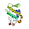
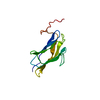
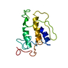
 Notechis scutatus scutatus (cobra)
 X-RAY DIFFRACTION /
  Authors
 Citation
 Structure visualization
 Downloads & links
 Other downloads


 PDBj
PDBj

 Assembly
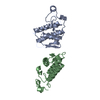
 Mass: 18.015 Da / Num. of mol.: 10 / Source method: isolated from a natural source / Formula: H2O
 Sample preparation
 Processing