 Movie
Movie Controller

[English] 日本語
 Yorodumi
- PDB-2no5: Crystal Structure analysis of a Dehalogenase with intermediate complex -
+ Open data

- Basic information
| Entry | Database: PDB / ID: 2no5 | ||||||
|---|---|---|---|---|---|---|---|
| Title | Crystal Structure analysis of a Dehalogenase with intermediate complex | ||||||
 Components Components | (S)-2-haloacid dehalogenase IVA | ||||||
 Keywords Keywords | HYDROLASE / Haloacid dehalogenase / Dehalogenase / HAD Superfamily / Intermediate structure | ||||||
| Function / homology |  Function and homology information Function and homology information | ||||||
| Biological species |  Burkholderia cepacia (bacteria) Burkholderia cepacia (bacteria) | ||||||
| Method |  X-RAY DIFFRACTION / FOURIER SYNTHESIS / Resolution: 2.6 Å X-RAY DIFFRACTION / FOURIER SYNTHESIS / Resolution: 2.6 Å | ||||||
 Authors Authors | Schmidberger, J.W. / Wilce, M.C.J. | ||||||
 Citation Citation | Journal: J.Mol.Biol. / Year: 2007 Title: Crystal structures of the substrate free-enzyme, and reaction intermediate of the HAD superfamily member, haloacid dehalogenase DehIVa from Burkholderia cepacia MBA4 Authors: Schmidberger, J.W. / Wilce, J.A. / Tsang, J.S.H. / Wilce, M.C.J. | ||||||
| History |
|
- Structure visualization
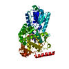
| Structure viewer | Molecule: MolmilJmol/JSmol |
|---|
- Downloads & links
-Download
| PDBx/mmCIF format | 2no5.cif.gz | 107.4 KB | Display | PDBx/mmCIF format |
|---|---|---|---|---|
| PDB format | pdb2no5.ent.gz | 82.9 KB | Display | PDB format |
| PDBx/mmJSON format | 2no5.json.gz | Tree view | PDBx/mmJSON format | |
| Others |  Other downloads Other downloads |
-Validation report
| Summary document | 2no5_validation.pdf.gz | 484.8 KB | Display | wwPDB validaton report |
|---|---|---|---|---|
| Full document | 2no5_full_validation.pdf.gz | 496 KB | Display | |
| Data in XML | 2no5_validation.xml.gz | 21.7 KB | Display | |
| Data in CIF | 2no5_validation.cif.gz | 28.9 KB | Display | |
| Arichive directory | https://data.pdbj.org/pub/pdb/validation_reports/no/2no5ftp://data.pdbj.org/pub/pdb/validation_reports/no/2no5 | HTTPS FTP |
-Related structure data
| Related structure data |  2no4SC S: Starting model for refinement C: citing same article ( |
|---|---|
| Similar structure data |


-Links
 PDBj
PDBj

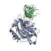
- Assembly
| Deposited unit | 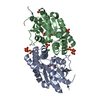
| ||||||||
|---|---|---|---|---|---|---|---|---|---|
| 1 |
| ||||||||
| 2 | 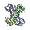
| ||||||||
| Unit cell |
| ||||||||
| Components on special symmetry positions |
| ||||||||
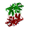
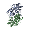
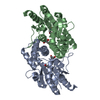
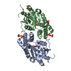
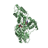
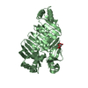
| Details | The structure is a Functional homodimer in the ASU. Asp11 on each chain (A and B) is covalently bound to a reaction intermediate, of the substrate L-2-monochloroprapanate. |
-Components
| #1: Protein | Mass: 27206.734 Da / Num. of mol.: 2 Source method: isolated from a genetically manipulated source Source: (gene. exp.) Burkholderia cepacia (bacteria) / Strain: MBA4 / Plasmid: pHKU201 / Production host: #2: Chemical | ChemComp-CL / |   Mass: 35.453 Da / Num. of mol.: 1 / Source method: obtained synthetically / Formula: Cl Mass: 35.453 Da / Num. of mol.: 1 / Source method: obtained synthetically / Formula: Cl#3: Chemical | ChemComp-SO4 /   Mass: 96.063 Da / Num. of mol.: 13 / Source method: obtained synthetically / Formula: SO4 Mass: 96.063 Da / Num. of mol.: 13 / Source method: obtained synthetically / Formula: SO4#4: Chemical | 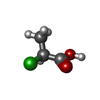  Mass: 108.524 Da / Num. of mol.: 3 / Source method: obtained synthetically / Formula: C3H5ClO2 Mass: 108.524 Da / Num. of mol.: 3 / Source method: obtained synthetically / Formula: C3H5ClO2#5: Water | ChemComp-HOH / |  Mass: 18.015 Da / Num. of mol.: 89 / Source method: isolated from a natural source / Formula: H2O Mass: 18.015 Da / Num. of mol.: 89 / Source method: isolated from a natural source / Formula: H2O |
|---|
-Experimental details
-Experiment
| Experiment | Method: X-RAY DIFFRACTION / Number of used crystals: 1 |
|---|
- Sample preparation
| Crystal | Density Matthews: 4.01 Å3/Da / Density % sol: 69.35 % |
|---|---|
| Crystal grow | Temperature: 293 K / Method: vapor diffusion, hanging drop / pH: 4.8 Details: 20% PEG 4000, 0.65M Ammonium Sulphate, pH 4.8, VAPOR DIFFUSION, HANGING DROP, temperature 293K |
-Data collection
| Diffraction | Mean temperature: 100 K |
|---|---|
| Diffraction source | Source: ROTATING ANODE / Type: RIGAKU / Wavelength: 1.5418 Å |
| Detector | Type: MAR scanner 345 mm plate / Detector: IMAGE PLATE / Date: Jul 16, 2004 / Details: Osmic mirrors |
| Radiation | Monochromator: osmic mirrors / Protocol: SINGLE WAVELENGTH / Monochromatic (M) / Laue (L): M / Scattering type: x-ray |
| Radiation wavelength | Wavelength: 1.5418 Å / Relative weight: 1 |
| Reflection | Resolution: 2.6→29.24 Å / Num. all: 26826 / Num. obs: 26322 / % possible obs: 99.9 % / Observed criterion σ(F): 1 / Redundancy: 3.49 % / Biso Wilson estimate: 55.1 Å2 / Rmerge(I) obs: 0.133 / Net I/σ(I): 10.13 |
| Reflection shell | Resolution: 2.58→2.67 Å / Redundancy: 3.11 % / Mean I/σ(I) obs: 1.41 / Num. unique all: 1919 / % possible all: 99.9 |
- Processing
| Software |
| ||||||||||||||||||||||||||||||||||||||||||||||||||||||||||||||||||||||||||||||||||||||||||
|---|---|---|---|---|---|---|---|---|---|---|---|---|---|---|---|---|---|---|---|---|---|---|---|---|---|---|---|---|---|---|---|---|---|---|---|---|---|---|---|---|---|---|---|---|---|---|---|---|---|---|---|---|---|---|---|---|---|---|---|---|---|---|---|---|---|---|---|---|---|---|---|---|---|---|---|---|---|---|---|---|---|---|---|---|---|---|---|---|---|---|---|
| Refinement | Method to determine structure: FOURIER SYNTHESIS Starting model: 2NO4 Resolution: 2.6→29.24 Å / Cor.coef. Fo:Fc: 0.938 / Cor.coef. Fo:Fc free: 0.899 / SU B: 8.856 / SU ML: 0.189 / Cross valid method: THROUGHOUT / σ(F): 0 / ESU R: 0.324 / ESU R Free: 0.261 / Stereochemistry target values: MAXIMUM LIKELIHOOD / Details: HYDROGENS HAVE BEEN ADDED IN THE RIDING POSITIONS
| ||||||||||||||||||||||||||||||||||||||||||||||||||||||||||||||||||||||||||||||||||||||||||
| Solvent computation | Ion probe radii: 0.8 Å / Shrinkage radii: 0.8 Å / VDW probe radii: 1.2 Å / Solvent model: MASK | ||||||||||||||||||||||||||||||||||||||||||||||||||||||||||||||||||||||||||||||||||||||||||
| Displacement parameters | Biso mean: 40.139 Å2
| ||||||||||||||||||||||||||||||||||||||||||||||||||||||||||||||||||||||||||||||||||||||||||
| Refinement step | Cycle: LAST / Resolution: 2.6→29.24 Å
| ||||||||||||||||||||||||||||||||||||||||||||||||||||||||||||||||||||||||||||||||||||||||||
| Refine LS restraints |
| ||||||||||||||||||||||||||||||||||||||||||||||||||||||||||||||||||||||||||||||||||||||||||
| LS refinement shell | Resolution: 2.599→2.666 Å / Total num. of bins used: 20
|
