 Movie
Movie Controller

[English] 日本語
 Yorodumi
- PDB-2nmn: Crystal structure of human galectin-3 carbohydrate-recognising do... -
+ Open data

- Basic information
| Entry | Database: PDB / ID: 2nmn | |||||||||
|---|---|---|---|---|---|---|---|---|---|---|
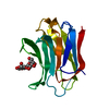
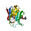
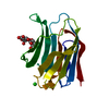
| Title | Crystal structure of human galectin-3 carbohydrate-recognising domain at 2.45 angstrom resolution | |||||||||
 Components Components | Galectin-3 | |||||||||
 Keywords Keywords | SUGAR BINDING PROTEIN / beta sandwich | |||||||||
| Function / homology |  Function and homology information Function and homology informationnegative regulation of NK T cell activation / negative regulation of immunological synapse formation / negative regulation of T cell activation via T cell receptor contact with antigen bound to MHC molecule on antigen presenting cell / disaccharide binding / RUNX2 regulates genes involved in differentiation of myeloid cells / regulation of T cell apoptotic process / mononuclear cell migration / receptor ligand inhibitor activity / negative regulation of endocytosis / positive regulation of mononuclear cell migration ...negative regulation of NK T cell activation / negative regulation of immunological synapse formation / negative regulation of T cell activation via T cell receptor contact with antigen bound to MHC molecule on antigen presenting cell / disaccharide binding / RUNX2 regulates genes involved in differentiation of myeloid cells / regulation of T cell apoptotic process / mononuclear cell migration / receptor ligand inhibitor activity / negative regulation of endocytosis / positive regulation of mononuclear cell migration / IgE binding / eosinophil chemotaxis / regulation of extrinsic apoptotic signaling pathway via death domain receptors / RUNX1 regulates transcription of genes involved in differentiation of myeloid cells / protein phosphatase inhibitor activity / positive chemotaxis / chemoattractant activity / positive regulation of calcium ion import / regulation of T cell proliferation / macrophage chemotaxis / Advanced glycosylation endproduct receptor signaling / monocyte chemotaxis / immunological synapse / ficolin-1-rich granule membrane / negative regulation of T cell receptor signaling pathway / neutrophil chemotaxis / epithelial cell differentiation / laminin binding / RNA splicing / secretory granule membrane / negative regulation of extrinsic apoptotic signaling pathway / positive regulation of protein localization to plasma membrane / spliceosomal complex / molecular condensate scaffold activity / positive regulation of protein-containing complex assembly / mRNA processing / carbohydrate binding / extracellular matrix / protein phosphatase binding / mitochondrial inner membrane / innate immune response / Neutrophil degranulation / cell surface / : / RNA binding / extracellular exosome / extracellular region / nucleoplasm / membrane / nucleus / plasma membrane / cytoplasm / cytosol Similarity search - Function | |||||||||
| Biological species |  Homo sapiens (human) Homo sapiens (human) | |||||||||
| Method |  X-RAY DIFFRACTION / FOURIER SYNTHESIS / Resolution: 2.45 Å X-RAY DIFFRACTION / FOURIER SYNTHESIS / Resolution: 2.45 Å | |||||||||
 Authors Authors | Blanchard, H. / Collins, P.M. | |||||||||
 Citation Citation | Journal: Acta Crystallogr.,Sect.D / Year: 2007 Title: Slow diffusion of lactose out of galectin-3 crystals monitored by X-ray crystallography: possible implications for ligand-exchange protocols. Authors: Collins, P.M. / Hidari, K.I. / Blanchard, H. | |||||||||
| History |
| |||||||||
| Remark 300 | BIOMOLECULE: 1 This entry contains the crystallographic asymmetric unit which consists of 1 chain(s) ...BIOMOLECULE: 1 This entry contains the crystallographic asymmetric unit which consists of 1 chain(s). the biological molecule(s) is expected to be full-length galectin-3 which consists of N-terminal domain and the CRD domain. see remark 350 for information on generating the biological molecule(s) of CRD domain. |
- Structure visualization
| Structure viewer | Molecule: MolmilJmol/JSmol |
|---|
- Downloads & links
-Download
| PDBx/mmCIF format | 2nmn.cif.gz | 44 KB | Display | PDBx/mmCIF format |
|---|---|---|---|---|
| PDB format | pdb2nmn.ent.gz | 28.9 KB | Display | PDB format |
| PDBx/mmJSON format | 2nmn.json.gz | Tree view | PDBx/mmJSON format | |
| Others |  Other downloads Other downloads |
-Validation report
| Arichive directory | https://data.pdbj.org/pub/pdb/validation_reports/nm/2nmnftp://data.pdbj.org/pub/pdb/validation_reports/nm/2nmn | HTTPS FTP |
|---|
-Related structure data
| Related structure data | 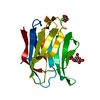 2nmoC 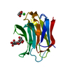 2nn8C 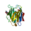 1a3kS C: citing same article ( S: Starting model for refinement |
|---|---|
| Similar structure data |


-Links
 PDBj
PDBj


- Assembly
| Deposited unit | 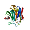
| ||||||||
|---|---|---|---|---|---|---|---|---|---|
| 1 |
| ||||||||
| Unit cell |
| ||||||||
| Details | The biological unit is a monomer of the Galectin-3 Carbohydrate-recognition domain. The crystallographic asymmetric unit contains one molecule of the Galectin-3 carbohydrate-recognition domain. |
-Components
| #1: Protein | Mass: 15701.049 Da / Num. of mol.: 1 / Fragment: Galectin-3 CRD domain, Residues 113-250 Source method: isolated from a genetically manipulated source Source: (gene. exp.) Homo sapiens (human) / Plasmid: pET-3a / Species (production host): Escherichia coli / Production host:  |
|---|---|
| #2: Polysaccharide | beta-D-galactopyranose-(1-4)-beta-D-glucopyranose / beta-lactose  Source method: isolated from a genetically manipulated source Details: oligosaccharide / References: beta-lactose |
| #3: Water | ChemComp-HOH /  Mass: 18.015 Da / Num. of mol.: 54 / Source method: isolated from a natural source / Formula: H2O Mass: 18.015 Da / Num. of mol.: 54 / Source method: isolated from a natural source / Formula: H2O |
-Experimental details
-Experiment
| Experiment | Method: X-RAY DIFFRACTION / Number of used crystals: 1 |
|---|
- Sample preparation
| Crystal | Density Matthews: 2.21 Å3/Da / Density % sol: 44.23 % |
|---|---|
| Crystal grow | Temperature: 293 K / Method: vapor diffusion, hanging drop / pH: 7 Details: 31% PEG 6000, 100mM MgCl2, 8mM beta-mercaptoethanol, 100mM Tris-HCL, pH 7.0, VAPOR DIFFUSION, HANGING DROP, temperature 293K |
-Data collection
| Diffraction | Mean temperature: 295 K |
|---|---|
| Diffraction source | Source: ROTATING ANODE / Type: MACSCIENCE / Wavelength: 1.5418 |
| Detector | Type: BRUKER SMART 6000 / Detector: CCD / Date: Jul 14, 2005 / Details: MIRRORS |
| Radiation | Monochromator: MIRRORS / Protocol: SINGLE WAVELENGTH / Monochromatic (M) / Laue (L): M / Scattering type: x-ray |
| Radiation wavelength | Wavelength: 1.5418 Å / Relative weight: 1 |
| Reflection | Resolution: 2.45→58 Å / Num. obs: 5400 / % possible obs: 98.4 % / Redundancy: 3.7 % / Rmerge(I) obs: 0.048 / Net I/σ(I): 11.4 |
| Reflection shell | Resolution: 2.45→2.56 Å / Rmerge(I) obs: 0.187 / Mean I/σ(I) obs: 4.6 / % possible all: 84.1 |
- Processing
| Software |
| ||||||||||||||||||||||||||||||||||||||||||||||||||||||||||||||||||||||||||||||||||||||||||||||||||||||||||||||||||||||||||||||||||||||||||||||||||||||||||||||||||||||||||
|---|---|---|---|---|---|---|---|---|---|---|---|---|---|---|---|---|---|---|---|---|---|---|---|---|---|---|---|---|---|---|---|---|---|---|---|---|---|---|---|---|---|---|---|---|---|---|---|---|---|---|---|---|---|---|---|---|---|---|---|---|---|---|---|---|---|---|---|---|---|---|---|---|---|---|---|---|---|---|---|---|---|---|---|---|---|---|---|---|---|---|---|---|---|---|---|---|---|---|---|---|---|---|---|---|---|---|---|---|---|---|---|---|---|---|---|---|---|---|---|---|---|---|---|---|---|---|---|---|---|---|---|---|---|---|---|---|---|---|---|---|---|---|---|---|---|---|---|---|---|---|---|---|---|---|---|---|---|---|---|---|---|---|---|---|---|---|---|---|---|---|---|
| Refinement | Method to determine structure: FOURIER SYNTHESIS Starting model: PDB ENTRY 1A3K Resolution: 2.45→42.99 Å / Cor.coef. Fo:Fc: 0.958 / Cor.coef. Fo:Fc free: 0.903 / SU B: 8.843 / SU ML: 0.191 / Cross valid method: THROUGHOUT / ESU R: 0.708 / ESU R Free: 0.285 / Stereochemistry target values: MAXIMUM LIKELIHOOD / Details: HYDROGENS HAVE BEEN ADDED IN THE RIDING POSITIONS
| ||||||||||||||||||||||||||||||||||||||||||||||||||||||||||||||||||||||||||||||||||||||||||||||||||||||||||||||||||||||||||||||||||||||||||||||||||||||||||||||||||||||||||
| Solvent computation | Ion probe radii: 0.8 Å / Shrinkage radii: 0.8 Å / VDW probe radii: 1.4 Å / Solvent model: MASK | ||||||||||||||||||||||||||||||||||||||||||||||||||||||||||||||||||||||||||||||||||||||||||||||||||||||||||||||||||||||||||||||||||||||||||||||||||||||||||||||||||||||||||
| Displacement parameters | Biso mean: 23.831 Å2
| ||||||||||||||||||||||||||||||||||||||||||||||||||||||||||||||||||||||||||||||||||||||||||||||||||||||||||||||||||||||||||||||||||||||||||||||||||||||||||||||||||||||||||
| Refinement step | Cycle: LAST / Resolution: 2.45→42.99 Å
| ||||||||||||||||||||||||||||||||||||||||||||||||||||||||||||||||||||||||||||||||||||||||||||||||||||||||||||||||||||||||||||||||||||||||||||||||||||||||||||||||||||||||||
| Refine LS restraints |
| ||||||||||||||||||||||||||||||||||||||||||||||||||||||||||||||||||||||||||||||||||||||||||||||||||||||||||||||||||||||||||||||||||||||||||||||||||||||||||||||||||||||||||
| LS refinement shell | Resolution: 2.446→2.509 Å / Total num. of bins used: 20
|
