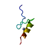
 Movie
Movie Controller


+ Open data

- Basic information
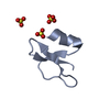
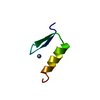
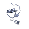
| Entry | Database: PDB / ID: 2nlh | ||||||
|---|---|---|---|---|---|---|---|
| Title | Human beta-defensin-1 (Mutant GLN24ALA) | ||||||
 Components Components | Beta-defensin 1 | ||||||
 Keywords Keywords | ANTIMICROBIAL PROTEIN / antimicrobial / chemotactic / defensin / mutant | ||||||
| Function / homology |  Function and homology information Function and homology informationpositive regulation of flagellated sperm motility involved in capacitation / CCR6 chemokine receptor binding / microvesicle / Beta defensins / Defensins / innate immune response in mucosa / response to bacterium / calcium-mediated signaling / Golgi lumen / chemotaxis ...positive regulation of flagellated sperm motility involved in capacitation / CCR6 chemokine receptor binding / microvesicle / Beta defensins / Defensins / innate immune response in mucosa / response to bacterium / calcium-mediated signaling / Golgi lumen / chemotaxis / antimicrobial humoral immune response mediated by antimicrobial peptide / antibacterial humoral response / sperm midpiece / defense response to Gram-negative bacterium / defense response to bacterium / defense response to Gram-positive bacterium / immune response / G protein-coupled receptor signaling pathway / innate immune response / : / extracellular exosome / extracellular region / membrane / identical protein binding Similarity search - Function | ||||||
| Biological species |  Homo sapiens (human) Homo sapiens (human) | ||||||
| Method |  X-RAY DIFFRACTION / MOLECULAR REPLACEMENT / Resolution: 1.85 Å X-RAY DIFFRACTION / MOLECULAR REPLACEMENT / Resolution: 1.85 Å | ||||||
 Authors Authors | Lubkowski, J. / Pazgier, M. | ||||||
 Citation Citation | Journal: J.Biol.Chem. / Year: 2007 Title: Studies of the Biological Properties of Human beta-Defensin 1. Authors: Pazgier, M. / Prahl, A. / Hoover, D.M. / Lubkowski, J. | ||||||
| History |
|
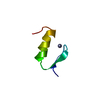
- Structure visualization
| Structure viewer | Molecule: MolmilJmol/JSmol |
|---|
- Downloads & links
-Download
| PDBx/mmCIF format | 2nlh.cif.gz | 45.9 KB | Display | PDBx/mmCIF format |
|---|---|---|---|---|
| PDB format | pdb2nlh.ent.gz | 33.1 KB | Display | PDB format |
| PDBx/mmJSON format | 2nlh.json.gz | Tree view | PDBx/mmJSON format | |
| Others |  Other downloads Other downloads |
-Validation report
| Arichive directory | https://data.pdbj.org/pub/pdb/validation_reports/nl/2nlhftp://data.pdbj.org/pub/pdb/validation_reports/nl/2nlh | HTTPS FTP |
|---|
-Related structure data
| Related structure data |  2nlbC  2nlcC 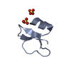 2nldC 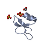 2nleC 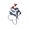 2nlfC 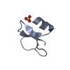 2nlgC 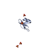 2nlpC  2nlqC  2nlsC 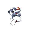 1ijvS C: citing same article ( S: Starting model for refinement |
|---|---|
| Similar structure data |


-Links
 PDBj
PDBj

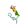
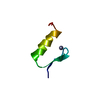
- Assembly
| Deposited unit | 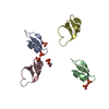
| ||||||||
|---|---|---|---|---|---|---|---|---|---|
| 1 | 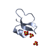
| ||||||||
| 2 | 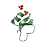
| ||||||||
| 3 | 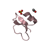
| ||||||||
| 4 | 
| ||||||||
| 5 | 
| ||||||||
| 6 | 
| ||||||||
| 7 | 
| ||||||||
| 8 | 
| ||||||||
| 9 | 
| ||||||||
| Unit cell |
| ||||||||
| Details | Biological assembly is a monomer |
-Components
| #1: Protein/peptide | Mass: 3883.547 Da / Num. of mol.: 4 / Fragment: human beta-defensins 1, residues 33-68 / Mutation: Q24A Source method: isolated from a genetically manipulated source Source: (gene. exp.) Homo sapiens (human) / Gene: DEFB1, BD1, HBD1 / Plasmid: pAED4 / Production host:  #2: Chemical | ChemComp-SO4 /   Mass: 96.063 Da / Num. of mol.: 6 / Source method: obtained synthetically / Formula: SO4 Mass: 96.063 Da / Num. of mol.: 6 / Source method: obtained synthetically / Formula: SO4#3: Chemical | ChemComp-ACT / |   Mass: 59.044 Da / Num. of mol.: 1 / Source method: obtained synthetically / Formula: C2H3O2 Mass: 59.044 Da / Num. of mol.: 1 / Source method: obtained synthetically / Formula: C2H3O2#4: Water | ChemComp-HOH / |  Mass: 18.015 Da / Num. of mol.: 196 / Source method: isolated from a natural source / Formula: H2O Mass: 18.015 Da / Num. of mol.: 196 / Source method: isolated from a natural source / Formula: H2OHas protein modification | Y | |
|---|
-Experimental details
-Experiment
| Experiment | Method: X-RAY DIFFRACTION / Number of used crystals: 1 |
|---|
- Sample preparation
| Crystal | Density Matthews: 2.23 Å3/Da / Density % sol: 44.95 % |
|---|---|
| Crystal grow | Temperature: 293 K / Method: vapor diffusion Details: PEG 4000, AMMONIUM SULFATE, vapor diffusion, temperature 293K |
-Data collection
| Diffraction | Mean temperature: 100 K |
|---|---|
| Diffraction source | Source: ROTATING ANODE / Type: RIGAKU RU200 / Wavelength: 1.54178 Å |
| Detector | Type: MAR scanner 345 mm plate / Detector: IMAGE PLATE / Date: Aug 15, 2005 / Details: Osmic mirrors |
| Radiation | Protocol: SINGLE WAVELENGTH / Monochromatic (M) / Laue (L): M / Scattering type: x-ray |
| Radiation wavelength | Wavelength: 1.54178 Å / Relative weight: 1 |
| Reflection | Resolution: 1.85→50 Å / Num. all: 12097 / Num. obs: 12097 / % possible obs: 100 % / Observed criterion σ(I): -3 / Redundancy: 7 % / Rmerge(I) obs: 0.066 / Χ2: 0.947 / Net I/σ(I): 27 |
| Reflection shell | Resolution: 1.85→1.92 Å / Redundancy: 6.3 % / Rmerge(I) obs: 0.506 / Mean I/σ(I) obs: 2.8 / Num. unique all: 1204 / Χ2: 0.698 / % possible all: 99.6 |
- Processing
| Software |
| ||||||||||||||||||||||||||||||||||||||||||||||||||||||||||||||||||||||||||||||||||||||||||
|---|---|---|---|---|---|---|---|---|---|---|---|---|---|---|---|---|---|---|---|---|---|---|---|---|---|---|---|---|---|---|---|---|---|---|---|---|---|---|---|---|---|---|---|---|---|---|---|---|---|---|---|---|---|---|---|---|---|---|---|---|---|---|---|---|---|---|---|---|---|---|---|---|---|---|---|---|---|---|---|---|---|---|---|---|---|---|---|---|---|---|---|
| Refinement | Method to determine structure: MOLECULAR REPLACEMENT Starting model: PDB ENTRY 1IJV Resolution: 1.85→30 Å / Cor.coef. Fo:Fc: 0.959 / Cor.coef. Fo:Fc free: 0.937 / SU B: 6.454 / SU ML: 0.109 / SU R Cruickshank DPI: 0.159 / Cross valid method: THROUGHOUT / ESU R Free: 0.154 / Stereochemistry target values: Engh & Huber
| ||||||||||||||||||||||||||||||||||||||||||||||||||||||||||||||||||||||||||||||||||||||||||
| Solvent computation | Ion probe radii: 0.8 Å / Shrinkage radii: 0.8 Å / VDW probe radii: 1.2 Å / Solvent model: MASK | ||||||||||||||||||||||||||||||||||||||||||||||||||||||||||||||||||||||||||||||||||||||||||
| Displacement parameters | Biso mean: 31.692 Å2
| ||||||||||||||||||||||||||||||||||||||||||||||||||||||||||||||||||||||||||||||||||||||||||
| Refinement step | Cycle: LAST / Resolution: 1.85→30 Å
| ||||||||||||||||||||||||||||||||||||||||||||||||||||||||||||||||||||||||||||||||||||||||||
| Refine LS restraints |
| ||||||||||||||||||||||||||||||||||||||||||||||||||||||||||||||||||||||||||||||||||||||||||
| LS refinement shell | Resolution: 1.849→1.897 Å / Total num. of bins used: 20
| ||||||||||||||||||||||||||||||||||||||||||||||||||||||||||||||||||||||||||||||||||||||||||
| Refinement TLS params. | Origin x: 7.9202 Å / Origin y: 7.7736 Å / Origin z: 20.3556 Å
| ||||||||||||||||||||||||||||||||||||||||||||||||||||||||||||||||||||||||||||||||||||||||||
| Refinement TLS group | Selection: ALL |
