- PDB-2nbq: NMR Structure of the C-Terminal Domain of human APOBEC3B -
+
Open data
ID or keywords:
Loading...
-
Basic information
Entry
Database: PDB / ID: 2nbq
Title
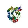
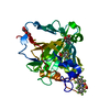
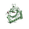
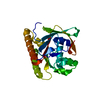
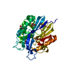
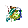
NMR Structure of the C-Terminal Domain of human APOBEC3B
Components
DNA dC->dU-editing enzyme APOBEC-3B
Keywords
HYDROLASE
Function / homology
Function and homology information
mRNA Editing: C to U Conversion / Formation of the Editosome / single-stranded DNA cytosine deaminase / negative regulation of single stranded viral RNA replication via double stranded DNA intermediate / DNA cytosine deamination / cytidine to uridine editing / clearance of foreign intracellular DNA / cytidine deaminase activity / transposable element silencing / P-body ...mRNA Editing: C to U Conversion / Formation of the Editosome / single-stranded DNA cytosine deaminase / negative regulation of single stranded viral RNA replication via double stranded DNA intermediate / DNA cytosine deamination / cytidine to uridine editing / clearance of foreign intracellular DNA / cytidine deaminase activity / transposable element silencing / P-body / defense response to virus / innate immune response / RNA binding / zinc ion binding / nucleoplasm / nucleus / cytoplasm Similarity search - Function
: / APOBEC3, cytosine deaminase domain / : / APOBEC/CMP deaminase, zinc-binding / Cytidine and deoxycytidylate deaminases zinc-binding region signature. / Cytidine and deoxycytidylate deaminase domain / Cytidine and deoxycytidylate deaminases domain profile. / Cytidine deaminase-like Similarity search - Domain/homology
Method: simulated annealing / Software ordinal: 1 Details: THE FLEXIBLE (HEXA-HISTIDINE TAG) RESIDUES WERE NOT INCLUDED IN THE STRUCTURE CALCULATIONS AND THUS NOT SHOWN IN THE COORDINATES.
NMR constraints
Hydrogen bond constraints total count: 168 / Protein chi angle constraints total count: 0 / Protein other angle constraints total count: 0 / Protein phi angle constraints total count: 155 / Protein psi angle constraints total count: 154
NMR representative
Selection criteria: lowest energy
NMR ensemble
Average torsion angle constraint violation: 0.397 ° Conformer selection criteria: structures with the lowest energy Conformers calculated total number: 256 / Conformers submitted total number: 30 / Maximum lower distance constraint violation: 0.5 Å / Maximum torsion angle constraint violation: 5 ° / Maximum upper distance constraint violation: 0.5 Å
NMR ensemble rms
Distance rms dev: 0.031 Å / Distance rms dev error: 0.002 Å
+
About Yorodumi
-
News
-
Feb 9, 2022. New format data for meta-information of EMDB entries
New format data for meta-information of EMDB entries
Version 3 of the EMDB header file is now the official format.
The previous official version 1.9 will be removed from the archive.
In the structure databanks used in Yorodumi, some data are registered as the other names, "COVID-19 virus" and "2019-nCoV". Here are the details of the virus and the list of structure data.
Jan 31, 2019. EMDB accession codes are about to change! (news from PDBe EMDB page)
EMDB accession codes are about to change! (news from PDBe EMDB page)
The allocation of 4 digits for EMDB accession codes will soon come to an end. Whilst these codes will remain in use, new EMDB accession codes will include an additional digit and will expand incrementally as the available range of codes is exhausted. The current 4-digit format prefixed with “EMD-” (i.e. EMD-XXXX) will advance to a 5-digit format (i.e. EMD-XXXXX), and so on. It is currently estimated that the 4-digit codes will be depleted around Spring 2019, at which point the 5-digit format will come into force.
The EM Navigator/Yorodumi systems omit the EMD- prefix.
Related info.:Q: What is EMD? / ID/Accession-code notation in Yorodumi/EM Navigator
Yorodumi is a browser for structure data from EMDB, PDB, SASBDB, etc.
This page is also the successor to EM Navigator detail page, and also detail information page/front-end page for Omokage search.
The word "yorodu" (or yorozu) is an old Japanese word meaning "ten thousand". "mi" (miru) is to see.
Related info.:EMDB / PDB / SASBDB / Comparison of 3 databanks / Yorodumi Search / Aug 31, 2016. New EM Navigator & Yorodumi / Yorodumi Papers / Jmol/JSmol / Function and homology information / Changes in new EM Navigator and Yorodumi
 Movie
Movie Controller


 Open data
 Basic information
 Components
 Keywords
 Function and homology information
 Homo sapiens (human)
 Authors
 Citation
 Structure visualization
 Downloads & links
 Other downloads
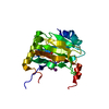


 PDBj
PDBj Assembly
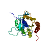


 Mass: 65.409 Da / Num. of mol.: 1 / Source method: obtained synthetically / Formula: Zn
 HSQC
HSQC Sample preparation
 Processing