[English] 日本語
Yorodumi
- PDB-2mzp: Structure and dynamics of the acidosis-resistant a162H mutant of ... -

+
Open data


ID or keywords:

Loading...

-
Basic information

Entry
Database: PDB / ID: 2mzp
TitleStructure and dynamics of the acidosis-resistant a162H mutant of the switch region of troponin I bound to the regulatory domain of troponin C
Components
  • Troponin C, slow skeletal and cardiac muscles
  • Troponin I, cardiac muscle
KeywordsCONTRACTILE PROTEIN / contraction regulation / contraction enhancenment / troponin / acidosis / Ca2+ binding
Function / homology
Function and homology information


regulation of systemic arterial blood pressure by ischemic conditions / troponin C binding / diaphragm contraction / regulation of ATP-dependent activity / regulation of muscle filament sliding speed / troponin T binding / cardiac myofibril / cardiac Troponin complex / troponin complex / regulation of muscle contraction ...regulation of systemic arterial blood pressure by ischemic conditions / troponin C binding / diaphragm contraction / regulation of ATP-dependent activity / regulation of muscle filament sliding speed / troponin T binding / cardiac myofibril / cardiac Troponin complex / troponin complex / regulation of muscle contraction / regulation of smooth muscle contraction / negative regulation of ATP-dependent activity / transition between fast and slow fiber / Striated Muscle Contraction / muscle filament sliding / response to metal ion / regulation of cardiac muscle contraction by calcium ion signaling / ventricular cardiac muscle tissue morphogenesis / heart contraction / troponin I binding / skeletal muscle contraction / calcium channel inhibitor activity / vasculogenesis / Ion homeostasis / cardiac muscle contraction / sarcomere / intracellular calcium ion homeostasis / calcium-dependent protein binding / actin filament binding / actin binding / heart development / protein domain specific binding / calcium ion binding / protein kinase binding / protein homodimerization activity / cytosol
Similarity search - Function
Troponin I residues 1-32 / Troponin I residues 1-32 / : / Troponin / Troponin domain superfamily / Troponin / EF-hand domain pair / : / EF-hand domain pair / EF-hand, calcium binding motif ...Troponin I residues 1-32 / Troponin I residues 1-32 / : / Troponin / Troponin domain superfamily / Troponin / EF-hand domain pair / : / EF-hand domain pair / EF-hand, calcium binding motif / EF-Hand 1, calcium-binding site / EF-hand calcium-binding domain. / EF-hand calcium-binding domain profile. / EF-hand domain / EF-hand domain pair
Similarity search - Domain/homology
Troponin I, cardiac muscle / Troponin C, slow skeletal and cardiac muscles
Similarity search - Component
Biological speciesHomo sapiens (human)
MethodSOLUTION NMR / simulated annealing
AuthorsPineda Sanabria, S.E. / Robertson, I.M. / Sykes, B.D.
CitationJournal: Biochemistry / Year: 2015
Title: Structure and Dynamics of the Acidosis-Resistant A162H Mutant of the Switch Region of Troponin I Bound to the Regulatory Domain of Troponin C.
Authors: Pineda-Sanabria, S.E. / Robertson, I.M. / Sykes, B.D.
History
DepositionFeb 20, 2015Deposition site: BMRB / Processing site: RCSB
Revision 1.0Jun 3, 2015Provider: repository / Type: Initial release
Revision 1.1Jul 1, 2015Group: Database references
Revision 1.2Jun 14, 2023Group: Data collection / Database references ...Data collection / Database references / Derived calculations / Other
Category: database_2 / pdbx_database_status ...database_2 / pdbx_database_status / pdbx_nmr_software / pdbx_struct_conn_angle / struct_conn / struct_ref_seq_dif / struct_site
Item: _database_2.pdbx_DOI / _database_2.pdbx_database_accession ..._database_2.pdbx_DOI / _database_2.pdbx_database_accession / _pdbx_database_status.status_code_nmr_data / _pdbx_nmr_software.name / _pdbx_struct_conn_angle.ptnr1_auth_comp_id / _pdbx_struct_conn_angle.ptnr1_auth_seq_id / _pdbx_struct_conn_angle.ptnr1_label_atom_id / _pdbx_struct_conn_angle.ptnr1_label_comp_id / _pdbx_struct_conn_angle.ptnr1_label_seq_id / _pdbx_struct_conn_angle.ptnr3_auth_comp_id / _pdbx_struct_conn_angle.ptnr3_auth_seq_id / _pdbx_struct_conn_angle.ptnr3_label_atom_id / _pdbx_struct_conn_angle.ptnr3_label_comp_id / _pdbx_struct_conn_angle.ptnr3_label_seq_id / _pdbx_struct_conn_angle.value / _struct_conn.pdbx_dist_value / _struct_conn.ptnr1_auth_comp_id / _struct_conn.ptnr1_auth_seq_id / _struct_conn.ptnr1_label_atom_id / _struct_conn.ptnr1_label_comp_id / _struct_conn.ptnr1_label_seq_id / _struct_ref_seq_dif.details / _struct_site.pdbx_auth_asym_id / _struct_site.pdbx_auth_comp_id / _struct_site.pdbx_auth_seq_id
Revision 1.3May 15, 2024Group: Data collection / Database references / Category: chem_comp_atom / chem_comp_bond / database_2 / Item: _database_2.pdbx_DOI

-
Structure visualization

Structure viewerMolecule:
MolmilJmol/JSmol

Downloads & links

-
Assembly

Deposited unit
C: Troponin C, slow skeletal and cardiac muscles
I: Troponin I, cardiac muscle
hetero molecules


Theoretical massNumber of molelcules
Total (without water)13,1833
Polymers13,1432
Non-polymers401
Water00
1


  • Idetical with deposited unit
  • defined by author&software
TypeNameSymmetry operationNumber
identity operation1_555x,y,z1
Buried area1190 Å2
ΔGint-11 kcal/mol
Surface area9020 Å2
MethodPISA
NMR ensembles
DataCriteria
Number of conformers (submitted / calculated)20 / 200structures with the lowest energy
RepresentativeModel #1closest to the average

-
Components

#1: Protein Troponin C, slow skeletal and cardiac muscles / TN-C


Mass: 10070.304 Da / Num. of mol.: 1 / Fragment: UNP residues 1-89
Source method: isolated from a genetically manipulated source
Source: (gene. exp.) Homo sapiens (human) / Gene: TNNC1, TNNC / Plasmid: pET3a-cNTnC / Production host: Escherichia coli (E. coli) / References: UniProt: P63316
#2: Protein/peptide Troponin I, cardiac muscle / Cardiac troponin I


Mass: 3072.637 Da / Num. of mol.: 1 / Fragment: UNP residues 145-171 / Mutation: A162H / Source method: obtained synthetically / Source: (synth.) Homo sapiens (human) / References: UniProt: P19429
#3: Chemical ChemComp-CA / CALCIUM ION


Mass: 40.078 Da / Num. of mol.: 1 / Source method: obtained synthetically / Formula: Ca

-
Experimental details

-
Experiment

Experiment
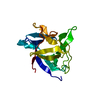
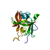
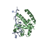
Method: SOLUTION NMR
Details: THE RELAXATION DATA WAS OBTAINED USING A CHIMERA PROTEIN THAT CONTAINS THE ACIDOSIS-RESISTANT A162H MUTANT OF THE SWITCH REGION OF TROPONIN I ATTACHED TO THE REGULATORY DOMAIN OF TROPONIN C ...Details: THE RELAXATION DATA WAS OBTAINED USING A CHIMERA PROTEIN THAT CONTAINS THE ACIDOSIS-RESISTANT A162H MUTANT OF THE SWITCH REGION OF TROPONIN I ATTACHED TO THE REGULATORY DOMAIN OF TROPONIN C THROUGH A FLEXIBLE LINKER. SEE PINEDA-SANABRIA ET AL (2014) ACS CHEM. BIOL. 9, 2121-2130 FOR REFERENCE.
NMR experiment
Conditions-IDExperiment-IDSolution-IDType
1112D 1H-15N HSQC
1223D HN(CA)CB
1323D CBCA(CO)NH
1422D 1H-13C HSQC
1523D H(CCO)NH
1623D C(CO)NH
1713D HNHA
1813D 1H-15N NOESY
1933D 1H-13C NOESY aliphatic
11042D DQF-COSY
11142D 1H-1H NOESY
11242D 1H-1H COSY
113313C-15N filtered TOCSY
114313C-15N filtered NOESY
1153Chmqcnoesy-Cfilt

-
Sample preparation

Details
Solution-IDContentsSolvent system
10.8-1 mM [U-15N] cNTnC, 3.2-4 mM cTnIA162H, 10 mM Calcium, 10 mM imidazole, 0.25 mM [U-2H] DSS, 10 mM DTT, 100 mM Potassium chloride, 95% H2O/5% D2O95% H2O/5% D2O
20.8-1 mM [U-13C; U-15N] cNTnC, 3.2-4 mM cTnIA162H, 10 mM Calcium, 10 mM imidazole, 0.25 mM [U-2H] DSS, 10 mM DTT, 100 mM Potassium chloride, 95% H2O/5% D2O95% H2O/5% D2O
30.8-1 mM [U-13C; U-15N] cNTnC, 3.2-4 mM cTnIA162H, 10 mM Calcium, 10 mM imidazole, 0.25 mM [U-2H] DSS, 10 mM DTT, 100 mM Potassium chloride, 100% D2O100% D2O
40.8-1 mM [U-15N] cNTnC, 3.2-4 mM cTnIA162H, 10 mM Calcium, 10 mM imidazole, 0.25 mM [U-2H] DSS, 10 mM DTT, 100 mM Potassium chloride, 100% D2O100% D2O
Sample
Conc. (mg/ml)UnitsComponentIsotopic labelingConc. range (mg/ml)Solution-ID
mMcNTnC-1[U-15N]0.8-11
mMcTnIA162H-23.2-41
10 mMCalcium-31
10 mMimidazole-41
0.25 mMDSS-5[U-2H]1
10 mMDTT-61
100 mMPotassium chloride-71
mMcNTnC-8[U-13C; U-15N]0.8-12
mMcTnIA162H-93.2-42
10 mMCalcium-102
10 mMimidazole-112
0.25 mMDSS-12[U-2H]2
10 mMDTT-132
100 mMPotassium chloride-142
mMcNTnC-15[U-13C; U-15N]0.8-13
mMcTnIA162H-163.2-43
10 mMCalcium-173
10 mMimidazole-183
0.25 mMDSS-19[U-2H]3
10 mMDTT-203
100 mMPotassium chloride-213
mMcNTnC-22[U-15N]0.8-14
mMcTnIA162H-233.2-44
10 mMCalcium-244
10 mMimidazole-254
0.25 mMDSS-26[U-2H]4
10 mMDTT-274
100 mMPotassium chloride-284
Sample conditionsIonic strength: 0.12 / pH: 6.1 / Pressure: ambient / Temperature: 303 K

-
NMR measurement

NMR spectrometer
TypeManufacturerModelField strength (MHz)Spectrometer-ID
Varian INOVAVarianINOVA5001
Varian INOVAVarianINOVA6002
Varian INOVAVarianINOVA8003

-
Processing

NMR software
NameVersionDeveloperClassification
VnmrJVariancollection
NMRPipeDelaglio, Grzesiek, Vuister, Zhu, Pfeifer and Baxprocessing
NMRDrawDelaglio, Grzesiek, Vuister, Zhu, Pfeifer and Baxprocessing
NMRViewJ8.2.33Johnson, One Moon Scientificchemical shift assignment
NMRViewJ8.2.33Johnson, One Moon Scientificdata analysis
NMRViewJ8.2.33Johnson, One Moon Scientificpeak picking
X-PLOR NIH2.35Schwieters, Kuszewski, Tjandra and Clorestructure solution
X-PLOR NIH2.35Schwieters, Kuszewski, Tjandra and Clorerefinement
PSVS1.5Bhattacharya and Montelionevalidation
RefinementMethod: simulated annealing / Software ordinal: 1
NMR constraintsNOE constraints total: 1282 / NOE intraresidue total count: 631 / NOE medium range total count: 303 / NOE sequential total count: 348
NMR representativeSelection criteria: closest to the average
NMR ensembleConformer selection criteria: structures with the lowest energy
Conformers calculated total number: 200 / Conformers submitted total number: 20 / Maximum lower distance constraint violation: 0.5 Å / Maximum upper distance constraint violation: 0.5 Å

+
About Yorodumi

-
News

-
Feb 9, 2022. New format data for meta-information of EMDB entries

New format data for meta-information of EMDB entries

  • Version 3 of the EMDB header file is now the official format.
  • The previous official version 1.9 will be removed from the archive.

Related info.:EMDB header

External links:wwPDB to switch to version 3 of the EMDB data model

-
Aug 12, 2020. Covid-19 info

Covid-19 info

URL: https://pdbj.org/emnavi/covid19.php

New page: Covid-19 featured information page in EM Navigator.

Related info.:Covid-19 info / Mar 5, 2020. Novel coronavirus structure data

+
Mar 5, 2020. Novel coronavirus structure data

Novel coronavirus structure data

Related info.:Yorodumi Speices / Aug 12, 2020. Covid-19 info

External links:COVID-19 featured content - PDBj / Molecule of the Month (242):Coronavirus Proteases

+
Jan 31, 2019. EMDB accession codes are about to change! (news from PDBe EMDB page)

EMDB accession codes are about to change! (news from PDBe EMDB page)

  • The allocation of 4 digits for EMDB accession codes will soon come to an end. Whilst these codes will remain in use, new EMDB accession codes will include an additional digit and will expand incrementally as the available range of codes is exhausted. The current 4-digit format prefixed with “EMD-” (i.e. EMD-XXXX) will advance to a 5-digit format (i.e. EMD-XXXXX), and so on. It is currently estimated that the 4-digit codes will be depleted around Spring 2019, at which point the 5-digit format will come into force.
  • The EM Navigator/Yorodumi systems omit the EMD- prefix.

Related info.:Q: What is EMD? / ID/Accession-code notation in Yorodumi/EM Navigator

External links:EMDB Accession Codes are Changing Soon! / Contact to PDBj

+
Jul 12, 2017. Major update of PDB

Major update of PDB

  • wwPDB released updated PDB data conforming to the new PDBx/mmCIF dictionary.
  • This is a major update changing the version number from 4 to 5, and with Remediation, in which all the entries are updated.
  • In this update, many items about electron microscopy experimental information are reorganized (e.g. em_software).
  • Now, EM Navigator and Yorodumi are based on the updated data.

External links:wwPDB Remediation / Enriched Model Files Conforming to OneDep Data Standards Now Available in the PDB FTP Archive

-
Yorodumi

Thousand views of thousand structures

  • Yorodumi is a browser for structure data from EMDB, PDB, SASBDB, etc.
  • This page is also the successor to EM Navigator detail page, and also detail information page/front-end page for Omokage search.
  • The word "yorodu" (or yorozu) is an old Japanese word meaning "ten thousand". "mi" (miru) is to see.

Related info.:EMDB / PDB / SASBDB / Comparison of 3 databanks / Yorodumi Search / Aug 31, 2016. New EM Navigator & Yorodumi / Yorodumi Papers / Jmol/JSmol / Function and homology information / Changes in new EM Navigator and Yorodumi

Read more