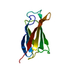
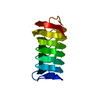
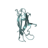
- PDB-2mb7: Solution structure of MBD3 methylcytosine binding domain -
+
Open data
ID or keywords:
Loading...
-
Basic information
Entry
Database: PDB / ID: 2mb7
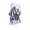
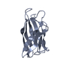
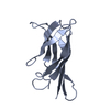
Title
Solution structure of MBD3 methylcytosine binding domain
Components
Methyl-CpG-binding domain protein 3
Keywords
TRANSCRIPTION / MBD3 / DNA methylation / NuRD / chromatin
Function / homology
Function and homology information
response to bisphenol A / ventricular cardiac muscle tissue development / NuRD complex / regulation of cell fate specification / regulation of stem cell differentiation / methyl-CpG binding / DNA methylation-dependent constitutive heterochromatin formation / RNA Polymerase I Transcription Initiation / embryonic organ development / Transcriptional regulation of brown and beige adipocyte differentiation by EBF2 ...response to bisphenol A / ventricular cardiac muscle tissue development / NuRD complex / regulation of cell fate specification / regulation of stem cell differentiation / methyl-CpG binding / DNA methylation-dependent constitutive heterochromatin formation / RNA Polymerase I Transcription Initiation / embryonic organ development / Transcriptional regulation of brown and beige adipocyte differentiation by EBF2 / Regulation of TP53 Activity through Acetylation / heterochromatin / Regulation of PTEN gene transcription / ERCC6 (CSB) and EHMT2 (G9a) positively regulate rRNA expression / Regulation of endogenous retroelements by KRAB-ZFP proteins / HDACs deacetylate histones / Regulation of endogenous retroelements by Piwi-interacting RNAs (piRNAs) / response to nutrient levels / response to estradiol / in utero embryonic development / Potential therapeutics for SARS / chromatin remodeling / negative regulation of DNA-templated transcription / positive regulation of DNA-templated transcription / chromatin / negative regulation of transcription by RNA polymerase II / protein-containing complex / DNA binding / nucleoplasm / nucleus / cytoplasm Similarity search - Function
Methyl-CpG binding protein 2/3, C-terminal domain / Methyl-CpG-binding domain protein 2/3, p55-binding region / C-terminal domain of methyl-CpG binding protein 2 and 3 / p55-binding region of Methyl-CpG-binding domain proteins MBD / Methyl-cpg-binding Protein 2; Chain A / Methyl-cpg-binding Protein 2; Chain A / Methyl-CpG binding domain / Methyl-CpG DNA binding / Methyl-CpG binding domain / Methyl-CpG-binding domain (MBD) profile. ...Methyl-CpG binding protein 2/3, C-terminal domain / Methyl-CpG-binding domain protein 2/3, p55-binding region / C-terminal domain of methyl-CpG binding protein 2 and 3 / p55-binding region of Methyl-CpG-binding domain proteins MBD / Methyl-cpg-binding Protein 2; Chain A / Methyl-cpg-binding Protein 2; Chain A / Methyl-CpG binding domain / Methyl-CpG DNA binding / Methyl-CpG binding domain / Methyl-CpG-binding domain (MBD) profile. / DNA-binding domain superfamily / 2-Layer Sandwich / Alpha Beta Similarity search - Domain/homology
NOE constraints total: 528 / NOE intraresidue total count: 102 / NOE long range total count: 160 / NOE medium range total count: 111 / NOE sequential total count: 155 / Protein chi angle constraints total count: 12 / Protein phi angle constraints total count: 54 / Protein psi angle constraints total count: 54
NMR representative
Selection criteria: lowest energy
NMR ensemble
Conformer selection criteria: structures with the lowest energy Conformers calculated total number: 50 / Conformers submitted total number: 20 / Maximum torsion angle constraint violation: 4.8 ° / Maximum upper distance constraint violation: 0.48 Å
NMR ensemble rms
Distance rms dev: 0.018 Å / Distance rms dev error: 0.003 Å
+
About Yorodumi
-
News
-
Feb 9, 2022. New format data for meta-information of EMDB entries
New format data for meta-information of EMDB entries
Version 3 of the EMDB header file is now the official format.
The previous official version 1.9 will be removed from the archive.
In the structure databanks used in Yorodumi, some data are registered as the other names, "COVID-19 virus" and "2019-nCoV". Here are the details of the virus and the list of structure data.
Jan 31, 2019. EMDB accession codes are about to change! (news from PDBe EMDB page)
EMDB accession codes are about to change! (news from PDBe EMDB page)
The allocation of 4 digits for EMDB accession codes will soon come to an end. Whilst these codes will remain in use, new EMDB accession codes will include an additional digit and will expand incrementally as the available range of codes is exhausted. The current 4-digit format prefixed with “EMD-” (i.e. EMD-XXXX) will advance to a 5-digit format (i.e. EMD-XXXXX), and so on. It is currently estimated that the 4-digit codes will be depleted around Spring 2019, at which point the 5-digit format will come into force.
The EM Navigator/Yorodumi systems omit the EMD- prefix.
Related info.:Q: What is EMD? / ID/Accession-code notation in Yorodumi/EM Navigator
Yorodumi is a browser for structure data from EMDB, PDB, SASBDB, etc.
This page is also the successor to EM Navigator detail page, and also detail information page/front-end page for Omokage search.
The word "yorodu" (or yorozu) is an old Japanese word meaning "ten thousand". "mi" (miru) is to see.
Related info.:EMDB / PDB / SASBDB / Comparison of 3 databanks / Yorodumi Search / Aug 31, 2016. New EM Navigator & Yorodumi / Yorodumi Papers / Jmol/JSmol / Function and homology information / Changes in new EM Navigator and Yorodumi
 Movie
Movie Controller


 Open data
 Basic information
 Components
 Keywords
 Function and homology information
 Homo sapiens (human)
 Authors
 Citation
 Structure visualization
 Downloads & links
 Other downloads


 PDBj
PDBj


 Assembly
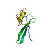

 HSQC
HSQC Sample preparation
 Processing