- PDB-2ma6: Solution NMR Structure of the RING finger domain from the Kip1 ub... -
+
Open data
ID or keywords:
Loading...
-
Basic information
Entry
Database: PDB / ID: 2ma6
Title
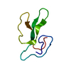
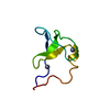
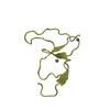
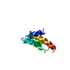
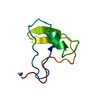
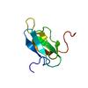
Solution NMR Structure of the RING finger domain from the Kip1 ubiquitination-promoting E3 complex protein 1 (KPC1/RNF123) from Homo sapiens, Northeast Structural Genomics Consortium (NESG) Target HR8700A
Components
E3 ubiquitin-protein ligase RNF123
Keywords
LIGASE / Structural Genomics / NORTHEAST STRUCTURAL GENOMICS CONSORTIUM / NESG / PSI-Biology / Protein Structure Initiative / Ring fing domain / Zinc binding protein
Function / homology
Function and homology information
: / protein maturation / RING-type E3 ubiquitin transferase / protein polyubiquitination / ubiquitin-protein transferase activity / ubiquitin protein ligase activity / Antigen processing: Ubiquitination & Proteasome degradation / ubiquitin-dependent protein catabolic process / nuclear membrane / Ub-specific processing proteases ...: / protein maturation / RING-type E3 ubiquitin transferase / protein polyubiquitination / ubiquitin-protein transferase activity / ubiquitin protein ligase activity / Antigen processing: Ubiquitination & Proteasome degradation / ubiquitin-dependent protein catabolic process / nuclear membrane / Ub-specific processing proteases / zinc ion binding / cytosol / cytoplasm Similarity search - Function
Mass: 65.409 Da / Num. of mol.: 2 / Source method: obtained synthetically / Formula: Zn
-
Experimental details
-
Experiment
Experiment
Method: SOLUTION NMR
NMR experiment
Conditions-ID
Experiment-ID
Solution-ID
Type
1
1
1
2D 1H-15N HSQC
1
2
1
2D 1H-13C HSQC aliphatic
1
3
1
2D 1H-13C HSQC aromatic CT
1
4
1
2D 1H-13C HSQC aromatic noCT
1
5
1
3D 1H-15N NOESY
1
6
1
3D 1H-15N NOESYNUS
1
7
1
3D 1H-13C NOESY aliphatic
1
8
1
3D 1H-13C NOESY aliphatic NUS
1
9
1
3D 1H-13C NOESY aromatic
1
10
2
2D 1H-13C HSQC aliphatic
1
11
1
3DC(CO)NH
1
12
1
3DH(CCO)NH
1
13
1
3D (H)CCH-COSY
1
14
3
2D 1H-15N HSQC
1
15
3
2D 1H-13C HSQC aliphatic
1
16
3
2D 1H-15N HSQC HIS
1
17
1
1DT1NSQCarray
1
18
1
1DT2NHSQCarray
1
19
1
3DCBCA(CO)NH
1
20
1
3D HN(CA)CB
1
21
1
3DHBHA(CO)NH
1
22
1
3D HNCO
1
23
1
2D 1H-15N HSQC NH2 only
1
24
2
4D (H)CCHNOESY
1
25
2
3D CCH-TOCSY
1
26
2
2D 1H-15N HSQC
1
27
1
2D 1H-15N HSQC
1
28
1
3D (H)CCH-TOCSY
-
Sample preparation
Details
Solution-ID
Contents
Solvent system
1
1.0 mM [U-15N] 5% 13C-fractional labeling HR8700A.005, 0.02 % NaN3, 10 mM DTT, 5 mM CaCL2, 100 mM NaCL, 1 x Proteinase Inhibitors, 20 mM MES pH 6.5, 10 % D2O, 50 uM DSS, 90% H2O/10% D2O
90% H2O/10% D2O
2
1.0 mM [U-100% 13C; U-100% 15N] HR8700A.005, 0.02 % NaN3, 10 mM DTT, 5 mM CaCL2, 100 mM NaCL, 1 x Proteinase Inhibitors, 20 mM MES pH 6.5, 100 % D2O, 50 uM DSS, 90% H2O/10% D2O
90% H2O/10% D2O
3
0.9 mM [U-100% 15N] 5% 13C-fractional labeling HR8700A.007, 0.02 % NaN3, 10 mM DTT, 5 mM CaCL2, 100 mM NaCL, 1 x Proteinase Inhibitors, 20 mM MES pH 6.5, 10 % D2O, 50 uM DSS, 90% H2O/10% D2O
90% H2O/10% D2O
Sample
Conc. (mg/ml)
Component
Isotopic labeling
Solution-ID
1.0mM
HR8700A.005-1
[U-15N] 5% 13C-fractional labeling
1
0.02 %
NaN3-2
1
10mM
DTT-3
1
5mM
CaCL2-4
1
100mM
NaCL-5
1
1 %
Proteinase Inhibitors-6
1
20mM
MES pH 6.5-7
1
10 %
D2O-8
1
50uM
DSS-9
1
1.0mM
HR8700A.005-10
[U-100% 13C; U-100% 15N]
2
0.02 %
NaN3-11
2
10mM
DTT-12
2
5mM
CaCL2-13
2
100mM
NaCL-14
2
1 %
Proteinase Inhibitors-15
2
20mM
MES pH 6.5-16
2
100 %
D2O-17
2
50uM
DSS-18
2
0.9mM
HR8700A.007-19
[U-100% 15N] 5% 13C-fractional labeling
3
0.02 %
NaN3-20
3
10mM
DTT-21
3
5mM
CaCL2-22
3
100mM
NaCL-23
3
1 %
Proteinase Inhibitors-24
3
20mM
MES pH 6.5-25
3
10 %
D2O-26
3
50uM
DSS-27
3
Sample conditions
pH: 6.5 / Pressure: ambient / Temperature: 298 K
-
NMR measurement
NMR spectrometer
Type
Manufacturer
Model
Field strength (MHz)
Spectrometer-ID
Bruker Avance II
Bruker
AVANCEII
850
1
Varian INOVA
Varian
INOVA
600
2
Bruker Avance III
Bruker
AVANCEIII
600
3
-
Processing
NMR software
Name
Version
Developer
Classification
CNS
1.3
Brunger, Adams, Clore, Gros, NilgesandRead
refinemen,structuresolution,geometryoptimization
CYANA
3
Guntert, MumenthalerandWuthrich
refinement,geometryoptimization,structuresolution
AutoStructure
2.1
Huang, Tejero, PowersandMontelione
dataanalysis,refinement
NMRPipe
NMRPipe-2008
Delaglio, Grzesiek, Vuister, Zhu, PfeiferandBax
processing
TopSpin
2.4
BrukerBiospin
collection
VnmrJ
vnmrj_1.1_D
Varian
collection
PINE
Bahrami, Markley, Assadi, andEghbalnia
chemicalshiftassignment
Sparky
3.113
Goddard
dataanalysis
TALOS+
Version1.2009.0721.18
Shen, Cornilescu, DelaglioandBax
geometryoptimization
PSVS
1.4
Bhattacharya, Montelione
structurevalidation
FMCGUI
fmcgui2.5_linux
AlexLemak, CherylArrowmith
refinement
CNS
refinement
Refinement
Method: simulated annealing, null / Software ordinal: 1 Details: CNS water refinement of Cyana structures via fmcGui, null
NMR representative
Selection criteria: lowest energy
NMR ensemble
Conformer selection criteria: structures with the lowest energy Conformers calculated total number: 100 / Conformers submitted total number: 20
+
About Yorodumi
-
News
-
Feb 9, 2022. New format data for meta-information of EMDB entries
New format data for meta-information of EMDB entries
Version 3 of the EMDB header file is now the official format.
The previous official version 1.9 will be removed from the archive.
In the structure databanks used in Yorodumi, some data are registered as the other names, "COVID-19 virus" and "2019-nCoV". Here are the details of the virus and the list of structure data.
Jan 31, 2019. EMDB accession codes are about to change! (news from PDBe EMDB page)
EMDB accession codes are about to change! (news from PDBe EMDB page)
The allocation of 4 digits for EMDB accession codes will soon come to an end. Whilst these codes will remain in use, new EMDB accession codes will include an additional digit and will expand incrementally as the available range of codes is exhausted. The current 4-digit format prefixed with “EMD-” (i.e. EMD-XXXX) will advance to a 5-digit format (i.e. EMD-XXXXX), and so on. It is currently estimated that the 4-digit codes will be depleted around Spring 2019, at which point the 5-digit format will come into force.
The EM Navigator/Yorodumi systems omit the EMD- prefix.
Related info.:Q: What is EMD? / ID/Accession-code notation in Yorodumi/EM Navigator
Yorodumi is a browser for structure data from EMDB, PDB, SASBDB, etc.
This page is also the successor to EM Navigator detail page, and also detail information page/front-end page for Omokage search.
The word "yorodu" (or yorozu) is an old Japanese word meaning "ten thousand". "mi" (miru) is to see.
Related info.:EMDB / PDB / SASBDB / Comparison of 3 databanks / Yorodumi Search / Aug 31, 2016. New EM Navigator & Yorodumi / Yorodumi Papers / Jmol/JSmol / Function and homology information / Changes in new EM Navigator and Yorodumi
 Movie
Movie Controller

 Yorodumi
 Open data
 Basic information
 Components
 Keywords
 Function and homology information
 Homo sapiens (human)
 Authors
 Citation
 Structure visualization
 Downloads & links
 Other downloads
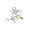

 PDBj
PDBj


 Assembly
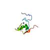


 Mass: 65.409 Da / Num. of mol.: 2 / Source method: obtained synthetically / Formula: Zn
 HSQC
HSQC Sample preparation
 Processing