 Movie
Movie Controller

[English] 日本語
 Yorodumi
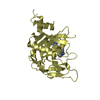
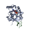
- PDB-2loy: Refined Miminal Constraint Solution NMR Structure of Translationa... -
+ Open data

- Basic information
| Entry | Database: PDB / ID: 2loy | ||||||
|---|---|---|---|---|---|---|---|
| Title | Refined Miminal Constraint Solution NMR Structure of Translationally-controlled tumor protein (TCTP) from Caenorhabditis elegans, Northeast Structural Genomics Consortium Target WR73 | ||||||
 Components Components | Translationally-controlled tumor protein homolog | ||||||
 Keywords Keywords | METAL BINDING PROTEIN / Structural Genomics / NORTHEAST STRUCTURAL GENOMICS CONSORTIUM (NESG) / PSI-Biology / Protein Structure Initiative | ||||||
| Function / homology |  Function and homology information Function and homology information | ||||||
| Biological species |  | ||||||
| Method | SOLUTION NMR / simulated annealing | ||||||
| Model details | lowest energy, model 1 | ||||||
 Authors Authors | Aramini, J.M. / Rossi, P. / Cort, J.R. / Lee, H. / Janjua, H. / Maglaqui, M. / Cooper, B. / Xiao, R. / Acton, T.B. / Everett, J.K. ...Aramini, J.M. / Rossi, P. / Cort, J.R. / Lee, H. / Janjua, H. / Maglaqui, M. / Cooper, B. / Xiao, R. / Acton, T.B. / Everett, J.K. / Montelione, G.T. / Northeast Structural Genomics Consortium (NESG) | ||||||
 Citation Citation | Journal: Proc.Natl.Acad.Sci.USA / Year: 2012 Title: Determination of solution structures of proteins up to 40 kDa using CS-Rosetta with sparse NMR data from deuterated samples. Authors: Lange, O.F. / Rossi, P. / Sgourakis, N.G. / Song, Y. / Lee, H.W. / Aramini, J.M. / Ertekin, A. / Xiao, R. / Acton, T.B. / Montelione, G.T. / Baker, D. | ||||||
| History |
|
- Structure visualization
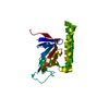
| Structure viewer | Molecule: MolmilJmol/JSmol |
|---|
- Downloads & links
-Download
| PDBx/mmCIF format | 2loy.cif.gz | 1.3 MB | Display | PDBx/mmCIF format |
|---|---|---|---|---|
| PDB format | pdb2loy.ent.gz | 1.1 MB | Display | PDB format |
| PDBx/mmJSON format | 2loy.json.gz | Tree view | PDBx/mmJSON format | |
| Others |  Other downloads Other downloads |
-Validation report
| Arichive directory | https://data.pdbj.org/pub/pdb/validation_reports/lo/2loyftp://data.pdbj.org/pub/pdb/validation_reports/lo/2loy | HTTPS FTP |
|---|
-Related structure data
| Related structure data | 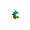 2kw5C  2kznC 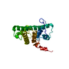 2lmdC 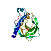 2lnuC 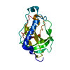 2lokC 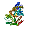 2mv0C C: citing same article ( |
|---|---|
| Similar structure data | |
| Other databases |

-Links
 PDBj
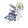
PDBj- Assembly
| Deposited unit | 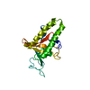
| |||||||||
|---|---|---|---|---|---|---|---|---|---|---|
| 1 |
| |||||||||
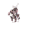
| NMR ensembles |
|
-Components
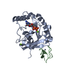
| #1: Protein | Mass: 21642.564 Da / Num. of mol.: 1 Source method: isolated from a genetically manipulated source Source: (gene. exp.)  |
|---|
-Experimental details
-Experiment
| Experiment | Method: SOLUTION NMR Details: Refinement of minimal constraint structure with residual dipolar couplings | ||||||||||||||||||||||||||||||||||||||||||||||||||||||||||||||||||||
|---|---|---|---|---|---|---|---|---|---|---|---|---|---|---|---|---|---|---|---|---|---|---|---|---|---|---|---|---|---|---|---|---|---|---|---|---|---|---|---|---|---|---|---|---|---|---|---|---|---|---|---|---|---|---|---|---|---|---|---|---|---|---|---|---|---|---|---|---|---|
| NMR experiment |
| ||||||||||||||||||||||||||||||||||||||||||||||||||||||||||||||||||||
| NMR details | Text: THE PROTEIN IS MONOMERIC AT 298 K BY 15N T1/T2 RELAXATION AND STATIC LIGHT SCATTERING. THE STRUCTURE IS A MINIMAL CONSTRAINT STRUCTURE DETERMINED USING TRIPLE RESONANCE NMR SPECTROSCOPY. ALL ...Text: THE PROTEIN IS MONOMERIC AT 298 K BY 15N T1/T2 RELAXATION AND STATIC LIGHT SCATTERING. THE STRUCTURE IS A MINIMAL CONSTRAINT STRUCTURE DETERMINED USING TRIPLE RESONANCE NMR SPECTROSCOPY. ALL NOESY DATA WERE ACQUIRED AT 800 MHZ USING A 5-MM CRYOPROBE. BACKBONE ASSIGNMENTS WERE MADE USING PINE, AND THE SIDE CHAIN METHYL ASSIGNMENTS WERE COMPLETED MANUALLY. AUTOMATIC NOESY ASSIGNMENTS WERE DETERMINED USING CYANA 3.0. BACKBONE (PHI/PSI) DIHEDRAL ANGLE CONSTRAINTS WERE OBTAINED FROM TALOSplus. FINAL STRUCTURE QUALITY FACTORS (FOR RESIDUE NUMBERS 1 TO 183, PSVS 1.4), WHERE ORDERED RESIDUES [S(PHI) + S(PSI) > 1.8] COMPRISE: 2-12,18-38,66-106,111-146,152-162,164-181: (A) RMSD (ORDERED RESIDUES): BB, 1.0, HEAVY ATOM, 1.6. (B) MOLPROBITY RAMACHANDRAN STATISTICS FOR ORDERED RESIDUES: MOST FAVORED, 96.8%, ALLOWED, 2.9%, DISALLOWED, 0.2%. (C) PROCHECK SCORES FOR ORDERED RESIDUES (RAW/Z-): PHI-PSI, -0.28/-0.79, ALL, -0.26/-1.54. (D) MOLPROBITY CLASH SCORE (RAW/Z-): 15.30/-1.10 (E) NUMBER OF CLOSE CONTACTS PER 20 MODELS: 40. (F) AGREEMENT WITH N-H RESIDUAL DIPOLAR COUPLINGS: CORRELATION COEFFICIENT (R): 0.985; Qrms: 0.171. THE C-TERMINAL HIS TAG RESIDUES OF THE PROTEIN (HHHHHH) WERE NOT INCLUDED IN THE STRUCTURE CALCULATIONS AND HAVE BEEN OMITTED FROM THIS DEPOSITION. COORDINATES FOR THE FOLLOWING RESIDUES ARE NOT WELL DETERMINED [S(PHI) + S(PSI) < 1.8]: 1,13-17,39-65,107-110,147-151,163,182-183. |
 HSQC
HSQC- Sample preparation
| Details |
| ||||||||||||||||||||||||||||||||||||||||||||||||||||||||||||||||||||||||||
|---|---|---|---|---|---|---|---|---|---|---|---|---|---|---|---|---|---|---|---|---|---|---|---|---|---|---|---|---|---|---|---|---|---|---|---|---|---|---|---|---|---|---|---|---|---|---|---|---|---|---|---|---|---|---|---|---|---|---|---|---|---|---|---|---|---|---|---|---|---|---|---|---|---|---|---|
| Sample |
|