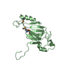
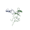
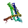
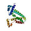
- PDB-2lev: Structure of the DNA complex of the C-Terminal domain of Ler -
+
Open data
ID or keywords:
Loading...
-
Basic information
Entry
Database: PDB / ID: 2lev
Title
Structure of the DNA complex of the C-Terminal domain of Ler
Components
DNA (5'-D(*CP*CP*TP*AP*TP*CP*AP*AP*TP*TP*AP*TP*CP*GP*C)-3')
DNA (5'-D(*GP*CP*GP*AP*TP*AP*AP*TP*TP*GP*AP*TP*AP*GP*G)-3')
Ler
Keywords
TRANSCRIPTION REGULATOR/DNA / TRANSCRIPTION REGULATOR-DNA complex / Arginine-minor-groove Recognition / Horizontal gene Transfer / Indirect Readout / Nucleoid associated Protein
Function / homology
Function and homology information
bent DNA binding / nucleoid / DNA-binding transcription repressor activity / minor groove of adenine-thymine-rich DNA binding / protein-DNA complex / transcription cis-regulatory region binding / cytosol Similarity search - Function
Histone-like protein H-NS, C-terminal domain / H-NS DNA Binding Protein / Histone-like protein H-NS, C-terminal domain superfamily / Histone-like protein H-NS, C-terminal domain / H-NS histone C-terminal domain / Domain in histone-like proteins of HNS family / Few Secondary Structures / Irregular Similarity search - Domain/homology
Mass: 6703.369 Da / Num. of mol.: 1 / Fragment: sequence database residues 56-102 Source method: isolated from a genetically manipulated source Source: (gene. exp.) Escherichia coli (E. coli) / Gene: ler / Production host: Escherichia coli (E. coli) / References: UniProt: E9N650, UniProt: Q7DB51*PLUS
#2: DNA chain
DNA (5'-D(*GP*CP*GP*AP*TP*AP*AP*TP*TP*GP*AP*TP*AP*GP*G)-3')
Mass: 4673.059 Da / Num. of mol.: 1 / Source method: obtained synthetically
#3: DNA chain
DNA (5'-D(*CP*CP*TP*AP*TP*CP*AP*AP*TP*TP*AP*TP*CP*GP*C)-3')
Mass: 4503.948 Da / Num. of mol.: 1 / Source method: obtained synthetically
-
Experimental details
-
Experiment
Experiment
Method
SOLUTION NMR
SOLUTION SCATTERING
NMR experiment
Conditions-ID
Experiment-ID
Solution-ID
Type
1
1
1
2D 1H-15N HSQC
1
2
3
2D 1H-13C HSQC aliphatic
1
3
1
3D HN(CA)CB
1
4
1
3DCBCA(CO)NH
1
5
1
3DH(CCO)NH
1
6
2
3D (H)CCH-TOCSY
1
7
1
3D HNHA
1
8
1
3D 1H-15N TOCSY
1
9
1
3D 1H-15N NOESY
1
10
1
3D 1H-13C NOESY aliphatic
1
11
2
3D 1H-13C NOESY aliphatic
1
12
2
2D 1H-1H (13C-ed) NOESY
1
13
1
2D 1H-1H NOESY
1
14
2
2D 1H-1H (13C-fil) NOESY
1
15
1
2D 1H-1H (15N/13C-fil) NOESY
1
16
1
3D 15N/13C-fil/13c-ed- NOESY
1
17
2
2D 1H-1H (13C-fil) TOCSY
-
Sample preparation
Details
Solution-ID
Contents
Solvent system
1
1 mM [U-100% 13C; U-100% 15N] CT-Ler, 2 mM LeeH A, 2 mM LeeH B, 20 mM sodium phosphate, 20 mM sodium chloride, 0.01 %(w/v) sodium azide, 0.2 mM EDTA, 90% H2O/10% D2O
90% H2O/10% D2O
2
1 mM [U-100% 13C; U-100% 15N] CT-Ler, 2 mM LeeH A, 2 mM LeeH B, 20 mM sodium phosphate, 150 mM sodium chloride, 0.01 %(w/v) sodium azide, 0.2 mM EDTA, 100% D2O
100% D2O
3
1 mM [U-10% 13C; U-100% 15N] CT-Ler, 2 mM LeeH A, 2 mM LeeH B, 20 mM sodium phosphate, 150 mM sodium chloride, 0.01 % (w/v) sodium azide, 0.2 mM EDTA, 90% H2O/10% D2O
 Movie
Movie Controller


 Open data
 Basic information
 Components
 Keywords
 Function and homology information
 SOLUTION SCATTERING / torsion angle dynamics, simulated annealing, molecular dynamics
  Authors
 Citation
 Structure visualization
 Downloads & links
 Other downloads


 PDBj
PDBj


 Assembly
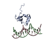
 Sample preparation
 Processing