[English] 日本語
 Yorodumi
- PDB-2lbf: Solution structure of the dimerization domain of human ribosomal ... -
+ Open data
- Basic information
| Entry | Database: PDB / ID: 2lbf | ||||||
|---|---|---|---|---|---|---|---|
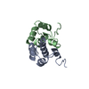
| Title | Solution structure of the dimerization domain of human ribosomal protein P1/P2 heterodimer | ||||||
 Components Components |
| ||||||
 Keywords Keywords | RIBOSOMAL PROTEIN / ribosome / stalk / P1/P2 | ||||||
| Function / homology |  Function and homology information Function and homology informationcytoplasmic translational elongation / Peptide chain elongation / Selenocysteine synthesis / Formation of a pool of free 40S subunits / translational elongation / Eukaryotic Translation Termination / SRP-dependent cotranslational protein targeting to membrane / Response of EIF2AK4 (GCN2) to amino acid deficiency / protein kinase activator activity / Viral mRNA Translation ...cytoplasmic translational elongation / Peptide chain elongation / Selenocysteine synthesis / Formation of a pool of free 40S subunits / translational elongation / Eukaryotic Translation Termination / SRP-dependent cotranslational protein targeting to membrane / Response of EIF2AK4 (GCN2) to amino acid deficiency / protein kinase activator activity / Viral mRNA Translation / Nonsense Mediated Decay (NMD) independent of the Exon Junction Complex (EJC) / GTP hydrolysis and joining of the 60S ribosomal subunit / ribonucleoprotein complex binding / L13a-mediated translational silencing of Ceruloplasmin expression / Major pathway of rRNA processing in the nucleolus and cytosol / Nonsense Mediated Decay (NMD) enhanced by the Exon Junction Complex (EJC) / cytosolic ribosome / Regulation of expression of SLITs and ROBOs / cytosolic large ribosomal subunit / cytoplasmic translation / structural constituent of ribosome / translation / focal adhesion / extracellular exosome / membrane / cytosol / cytoplasm Similarity search - Function | ||||||
| Biological species |  Homo sapiens (human) Homo sapiens (human) | ||||||
| Method | SOLUTION NMR / DGSA-distance geometry simulated annealing | ||||||
| Model details | closest to the average, model 1 | ||||||
 Authors Authors | Lee, K.-M. / Yu, C.W.-H. / Chiu, T.Y.-H. / Sze, K.-H. / Shaw, P.-C. / Wong, K.-B. | ||||||
 Citation Citation |  Journal: Nucleic Acids Res. / Year: 2011 Journal: Nucleic Acids Res. / Year: 2011Title: Solution structure of the dimerization domain of the eukaryotic stalk P1/P2 complex reveals the structural organization of eukaryotic stalk complex Authors: Lee, K.-M. / Yu, C.W.-H. / Chiu, T.Y.-H. / Sze, K.-H. / Shaw, P.-C. / Wong, K.-B. | ||||||
| History |
|
- Structure visualization
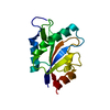
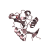
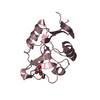
| Structure viewer | Molecule:  Molmil Molmil Jmol/JSmol Jmol/JSmol |
|---|
- Downloads & links
- Download
| PDBx/mmCIF format |  2lbf.cif.gz 2lbf.cif.gz | 397.4 KB | Display |  PDBx/mmCIF format PDBx/mmCIF format |
|---|---|---|---|---|
| PDB format |  pdb2lbf.ent.gz pdb2lbf.ent.gz | 331 KB | Display |  PDB format PDB format |
| PDBx/mmJSON format |  2lbf.json.gz 2lbf.json.gz | Tree view |  PDBx/mmJSON format PDBx/mmJSON format | |
| Others |  Other downloads Other downloads |
-Validation report
| Summary document |  2lbf_validation.pdf.gz 2lbf_validation.pdf.gz | 547.2 KB | Display |  wwPDB validaton report wwPDB validaton report |
|---|---|---|---|---|
| Full document |  2lbf_full_validation.pdf.gz 2lbf_full_validation.pdf.gz | 1.6 MB | Display | |
| Data in XML |  2lbf_validation.xml.gz 2lbf_validation.xml.gz | 175.7 KB | Display | |
| Data in CIF |  2lbf_validation.cif.gz 2lbf_validation.cif.gz | 166.9 KB | Display | |
| Arichive directory |  https://data.pdbj.org/pub/pdb/validation_reports/lb/2lbf https://data.pdbj.org/pub/pdb/validation_reports/lb/2lbf ftp://data.pdbj.org/pub/pdb/validation_reports/lb/2lbf ftp://data.pdbj.org/pub/pdb/validation_reports/lb/2lbf | HTTPS FTP |
-Related structure data
| Similar structure data | |
|---|---|
| Other databases |
- Links
Links
- Assembly
| Deposited unit | 
| |||||||||
|---|---|---|---|---|---|---|---|---|---|---|
| 1 |
| |||||||||
| NMR ensembles |
|
- Components
| #1: Protein | Mass: 7088.140 Da / Num. of mol.: 1 / Fragment: UNP residues 1-69 Source method: isolated from a genetically manipulated source Source: (gene. exp.)  Homo sapiens (human) / Gene: RPLP1, RRP1 / Production host: Homo sapiens (human) / Gene: RPLP1, RRP1 / Production host:  |
|---|---|
| #2: Protein | Mass: 7207.184 Da / Num. of mol.: 1 / Fragment: UNP residues 1-69 Source method: isolated from a genetically manipulated source Source: (gene. exp.)  Homo sapiens (human) / Gene: RPLP2, D11S2243E, RPP2 / Production host: Homo sapiens (human) / Gene: RPLP2, D11S2243E, RPP2 / Production host:  |
-Experimental details
-Experiment
| Experiment | Method: SOLUTION NMR |
|---|---|
| NMR experiment | Type: 2D 1H-15N  HSQC HSQC |
- Sample preparation
| Details | Contents: 1 mM [U-100% 13C; U-100% 15N] protein_1-1, 1 mM [U-100% 13C; U-100% 15N] protein_2-2, 90% H2O/10% D2O Solvent system: 90% H2O/10% D2O | ||||||||||||
|---|---|---|---|---|---|---|---|---|---|---|---|---|---|
| Sample |
| ||||||||||||
| Sample conditions | Ionic strength: 0.15 / pH: 6.5 / Pressure: ambient / Temperature: 298 K |
-NMR measurement
| NMR spectrometer | Type: Bruker Avance / Manufacturer: Bruker / Model: AVANCE / Field strength: 600 MHz |
|---|
- Processing
| NMR software |
| ||||||||||||
|---|---|---|---|---|---|---|---|---|---|---|---|---|---|
| Refinement | Method: DGSA-distance geometry simulated annealing / Software ordinal: 1 | ||||||||||||
| NMR representative | Selection criteria: closest to the average | ||||||||||||
| NMR ensemble | Conformer selection criteria: structures with the least restraint violations Conformers calculated total number: 100 / Conformers submitted total number: 10 / Representative conformer: 3 |
 Movie
Movie Controller


 PDBj
PDBj


