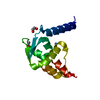
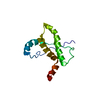
Method: simulated annealing / Software ordinal: 1 Details: The structure was determined using triple resonance NMR spectroscopy. Automated backbone assignments were made using Autoassign. Sidechain assignments were completed manually. Automated ...Details: The structure was determined using triple resonance NMR spectroscopy. Automated backbone assignments were made using Autoassign. Sidechain assignments were completed manually. Automated NOESY assignments were made using AutoStructure and structure solution was obtained using AutoStructure and CYANA-2.1. 150 structures were calculated and 20 best structures were refined in a shell of water using CNS. Initial dihedral angles were obtained using TALOS. The structure calculations were done including the N-terminal 6xHis tag. Resonance assignments were validated using AVS validation software. Final quality scores were determined using PSVS software. Ordered residues are defined as:11-13,18-54,62-77 . RMSD(ordered residuesall backbone aatoms 0.9A; All heavy atoms 1.4A; Ramachandran Statistics for all ordered residues: Most favoured 94.6%, additionally allowed region: 5.4%; Procheck scores for all ordered residues (Raw/Z) phi-psi 0.09/0.67; All dihedral angles 0.04/0.24; MolProbity clash score (Raw/Z) 13.00/-0.71. RPF scores for the goodness of fir of the structure to the NMR data: recall:0.933; Precision 0.918; F-measure 0.926 and final DP score: 0.768
NMR constraints
NOE constraints total: 712 / NOE intraresidue total count: 80 / NOE long range total count: 151 / NOE medium range total count: 236 / NOE sequential total count: 245
NMR representative
Selection criteria: lowest energy
NMR ensemble
Conformer selection criteria: structures with the lowest energy Conformers calculated total number: 150 / Conformers submitted total number: 20 / Maximum lower distance constraint violation: 0 Å / Maximum upper distance constraint violation: 0.1 Å
NMR ensemble rms
Distance rms dev: 0 Å
+
About Yorodumi
-
News
-
Feb 9, 2022. New format data for meta-information of EMDB entries
New format data for meta-information of EMDB entries
Version 3 of the EMDB header file is now the official format.
The previous official version 1.9 will be removed from the archive.
In the structure databanks used in Yorodumi, some data are registered as the other names, "COVID-19 virus" and "2019-nCoV". Here are the details of the virus and the list of structure data.
Jan 31, 2019. EMDB accession codes are about to change! (news from PDBe EMDB page)
EMDB accession codes are about to change! (news from PDBe EMDB page)
The allocation of 4 digits for EMDB accession codes will soon come to an end. Whilst these codes will remain in use, new EMDB accession codes will include an additional digit and will expand incrementally as the available range of codes is exhausted. The current 4-digit format prefixed with “EMD-” (i.e. EMD-XXXX) will advance to a 5-digit format (i.e. EMD-XXXXX), and so on. It is currently estimated that the 4-digit codes will be depleted around Spring 2019, at which point the 5-digit format will come into force.
The EM Navigator/Yorodumi systems omit the EMD- prefix.
Related info.:Q: What is EMD? / ID/Accession-code notation in Yorodumi/EM Navigator
Yorodumi is a browser for structure data from EMDB, PDB, SASBDB, etc.
This page is also the successor to EM Navigator detail page, and also detail information page/front-end page for Omokage search.
The word "yorodu" (or yorozu) is an old Japanese word meaning "ten thousand". "mi" (miru) is to see.
Related info.:EMDB / PDB / SASBDB / Comparison of 3 databanks / Yorodumi Search / Aug 31, 2016. New EM Navigator & Yorodumi / Yorodumi Papers / Jmol/JSmol / Function and homology information / Changes in new EM Navigator and Yorodumi
 Movie
Movie Controller

 Yorodumi
 Open data
 Basic information
 Components
 Keywords
 Function and homology information
 Homo sapiens (human)
 Authors
 Citation
 Structure visualization
 Downloads & links
 Other downloads
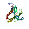


 PDBj
PDBj


 Assembly
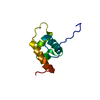

 HSQC
HSQC Sample preparation
 Processing