[English] 日本語
Yorodumi
- PDB-2k9y: EphA2 dimeric structure in the lipidic bicelle at pH 5.0 -

+
Open data


ID or keywords:

Loading...

-
Basic information

Entry
Database: PDB / ID: 2k9y
TitleEphA2 dimeric structure in the lipidic bicelle at pH 5.0
ComponentsEphrin type-A receptor 2
KeywordsTRANSFERASE / receptor tyrosine kinase / membrane protein / dimeric transmembrane domain / ephrin receptor / ATP-binding / Glycoprotein / Kinase / Membrane / Nucleotide-binding / Phosphoprotein / Polymorphism / Receptor / Transmembrane / Tyrosine-protein kinase
Function / homology
Function and homology information


notochord cell development / notochord formation / lens fiber cell morphogenesis / blood vessel endothelial cell proliferation involved in sprouting angiogenesis / negative regulation of lymphangiogenesis / axial mesoderm formation / pericyte cell differentiation / cAMP metabolic process / positive regulation of bicellular tight junction assembly / regulation of blood vessel endothelial cell migration ...notochord cell development / notochord formation / lens fiber cell morphogenesis / blood vessel endothelial cell proliferation involved in sprouting angiogenesis / negative regulation of lymphangiogenesis / axial mesoderm formation / pericyte cell differentiation / cAMP metabolic process / positive regulation of bicellular tight junction assembly / regulation of blood vessel endothelial cell migration / leading edge membrane / negative regulation of chemokine production / post-anal tail morphogenesis / bone remodeling / transmembrane-ephrin receptor activity / response to growth factor / tight junction / activation of GTPase activity / regulation of lamellipodium assembly / branching involved in mammary gland duct morphogenesis / EPH-Ephrin signaling / neural tube development / RND1 GTPase cycle / RND2 GTPase cycle / RND3 GTPase cycle / mammary gland epithelial cell proliferation / RHOV GTPase cycle / EPHA-mediated growth cone collapse / growth factor binding / regulation of cell adhesion mediated by integrin / lamellipodium membrane / RHOU GTPase cycle / RHOG GTPase cycle / EPH-ephrin mediated repulsion of cells / ephrin receptor signaling pathway / RAC2 GTPase cycle / RAC3 GTPase cycle / negative regulation of phosphatidylinositol 3-kinase/protein kinase B signal transduction / vasculogenesis / regulation of angiogenesis / keratinocyte differentiation / RAC1 GTPase cycle / transmembrane receptor protein tyrosine kinase activity / negative regulation of angiogenesis / phosphatidylinositol 3-kinase/protein kinase B signal transduction / regulation of ERK1 and ERK2 cascade / osteoclast differentiation / cell chemotaxis / skeletal system development / molecular function activator activity / protein localization to plasma membrane / cell motility / positive regulation of protein localization to plasma membrane / axon guidance / receptor protein-tyrosine kinase / ruffle membrane / osteoblast differentiation / intrinsic apoptotic signaling pathway in response to DNA damage / cell migration / lamellipodium / virus receptor activity / receptor complex / cell adhesion / defense response to Gram-positive bacterium / positive regulation of cell migration / cadherin binding / inflammatory response / phosphorylation / focal adhesion / dendrite / cell surface / ATP binding / plasma membrane
Similarity search - Function
Single helix bin / Ephrin type-A receptor 2, ligand binding domain / Ephrin receptor type-A /type-B / Ephrin receptor ligand binding domain / Tyrosine-protein kinase, receptor class V, conserved site / Ephrin receptor, transmembrane domain / Ephrin receptor ligand binding domain / Ephrin type-A receptor 2 transmembrane domain / Receptor tyrosine kinase class V signature 1. / Receptor tyrosine kinase class V signature 2. ...Single helix bin / Ephrin type-A receptor 2, ligand binding domain / Ephrin receptor type-A /type-B / Ephrin receptor ligand binding domain / Tyrosine-protein kinase, receptor class V, conserved site / Ephrin receptor, transmembrane domain / Ephrin receptor ligand binding domain / Ephrin type-A receptor 2 transmembrane domain / Receptor tyrosine kinase class V signature 1. / Receptor tyrosine kinase class V signature 2. / Eph receptor ligand-binding domain profile. / Ephrin receptor ligand binding domain / Putative ephrin-receptor like / SAM domain (Sterile alpha motif) / SAM domain profile. / Sterile alpha motif. / Sterile alpha motif domain / Sterile alpha motif/pointed domain superfamily / Growth factor receptor cysteine-rich domain superfamily / Fibronectin type III domain / Single alpha-helices involved in coiled-coils or other helix-helix interfaces / Fibronectin type 3 domain / Fibronectin type-III domain profile. / Galactose-binding-like domain superfamily / Fibronectin type III / Fibronectin type III superfamily / Tyrosine-protein kinase, catalytic domain / Tyrosine kinase, catalytic domain / Tyrosine protein kinases specific active-site signature. / Tyrosine-protein kinase, active site / Protein tyrosine and serine/threonine kinase / Serine-threonine/tyrosine-protein kinase, catalytic domain / Protein kinase, ATP binding site / Protein kinases ATP-binding region signature. / Immunoglobulin-like fold / Protein kinase domain profile. / Protein kinase domain / Protein kinase-like domain superfamily / Up-down Bundle / Mainly Alpha
Similarity search - Domain/homology
Ephrin type-A receptor 2
Similarity search - Component
Biological speciesHomo sapiens (human)
MethodSOLUTION NMR / TORSION ANGLE DYNAMICS, molecular dynamics, TORSION ANGLE DYNAMICS
AuthorsMayzel, M.L. / Bocharov, E.V. / Volynsky, P.E. / Arseniev, A.S.
CitationJournal: Biophys.J. / Year: 2010
Title: Left-Handed Dimer of EphA2 Transmembrane Domain: Helix Packing Diversity among Receptor Tyrosine Kinases
Authors: Bocharov, E.V. / Mayzel, M.L. / Volynsky, P.E. / Mineev, K.S. / Tkach, E.N. / Ermolyuk, Y.S. / Schulga, A.A. / Efremov, R.G. / Arseniev, A.S.
History
DepositionOct 27, 2008Deposition site: BMRB / Processing site: PDBJ
Revision 1.0Aug 11, 2009Provider: repository / Type: Initial release
Revision 1.1Jul 13, 2011Group: Version format compliance
Revision 1.2Mar 16, 2022Group: Database references / Derived calculations / Experimental preparation
Category: database_2 / pdbx_nmr_sample_details ...database_2 / pdbx_nmr_sample_details / pdbx_struct_assembly / pdbx_struct_assembly_prop / pdbx_struct_oper_list
Item: _database_2.pdbx_DOI / _database_2.pdbx_database_accession / _pdbx_nmr_sample_details.contents
Revision 1.3May 15, 2024Group: Data collection / Database references ...Data collection / Database references / Derived calculations / Source and taxonomy / Structure summary
Category: chem_comp_atom / chem_comp_bond ...chem_comp_atom / chem_comp_bond / database_2 / entity / entity_name_com / entity_src_gen / pdbx_struct_assembly_prop
Item: _database_2.pdbx_DOI / _entity.pdbx_ec ..._database_2.pdbx_DOI / _entity.pdbx_ec / _entity_src_gen.gene_src_common_name / _entity_src_gen.pdbx_beg_seq_num / _entity_src_gen.pdbx_end_seq_num / _entity_src_gen.pdbx_gene_src_gene / _entity_src_gen.pdbx_seq_type

-
Structure visualization

Structure viewerMolecule:
MolmilJmol/JSmol

Downloads & links

-
Assembly

Deposited unit
A: Ephrin type-A receptor 2
B: Ephrin type-A receptor 2


Theoretical massNumber of molelcules
Total (without water)8,5662
Polymers8,5662
Non-polymers00
Water543
1


  • Idetical with deposited unit
  • defined by author
TypeNameSymmetry operationNumber
identity operation1_5551
NMR ensembles
DataCriteria
Number of conformers (submitted / calculated)17 / 100structures with the least restraint violations
RepresentativeModel #1fewest violations

-
Components

#1: Protein/peptide Ephrin type-A receptor 2


Mass: 4283.052 Da / Num. of mol.: 2 / Fragment: EphA2_TM, residues 523-563
Source method: isolated from a genetically manipulated source
Source: (gene. exp.) Homo sapiens (human) / Gene: EPHA2 / Production host: Escherichia coli (E. coli) / References: UniProt: P29317
#2: Water ChemComp-HOH / water


Mass: 18.015 Da / Num. of mol.: 3 / Source method: isolated from a natural source / Formula: H2O

-
Experimental details

-
Experiment

ExperimentMethod: SOLUTION NMR
NMR experiment
Conditions-IDExperiment-IDSolution-IDType
1122D 1H-15N HSQC
1212D 1H-13C HSQC
1313D HNCO
1413D HNCA
1513D (H)CCH-TOCSY
1623D 1H-15N NOESY
1723D 1H-15N TOCSY
1813D 1H-13C NOESY
1923D HNHB
11023D HNHA
11122D 1H-1H NOESY
112313C F1-filtered/F3-edited-NOESY
113215N-T2
114215N-T1
115215N-NOE
11622D 1H-1H TOCSY

-
Sample preparation

Details
Solution-IDContentsSolvent system
13 mM [U-95% 13C; U-95% 15N] EphA2_TM 15N,13C, 96 mM [U-2H] DHPC, 24 mM [U-2H] DMPC, 1.5 mM NaN3, 1 mM EDTA, 10 mM phosphate buffer, 95% H2O/5% D2O95% H2O/5% D2O
23 mM [U-95% 15N] EphA2_TM 15N, 96 mM [U-2H] DHPC, 24 mM [U-2H] DMPC, 1.5 mM NaN3, 1 mM EDTA, 10 mM phosphate buffer, 90% H2O/10% D2O90% H2O/10% D2O
31.5 mM [U-95% 13C; U-95% 15N] EphA2_TM 15N,13C, 96 mM [U-2H] DHPC, 24 mM [U-2H] DMPC, 1.5 mM NaN3, 1 mM EDTA, 10 mM phosphate buffer, 1.5 mM EphA2_TM, 100% D2O100% D2O
Sample
Conc. (mg/ml)ComponentIsotopic labelingSolution-ID
3 mMEphA2_TM 15N,13C[U-95% 13C; U-95% 15N]1
96 mMDHPC[U-2H]1
24 mMDMPC[U-2H]1
1.5 mMNaN31
1 mMEDTA1
10 mMphosphate buffer1
3 mMEphA2_TM 15N[U-95% 15N]2
96 mMDHPC[U-2H]2
24 mMDMPC[U-2H]2
1.5 mMNaN32
1 mMEDTA2
10 mMphosphate buffer2
1.5 mMEphA2_TM 15N,13C[U-95% 13C; U-95% 15N]3
96 mMDHPC[U-2H]3
24 mMDMPC[U-2H]3
1.5 mMNaN33
1 mMEDTA3
10 mMphosphate buffer3
1.5 mMEphA2_TM3
Sample conditionsIonic strength: 10 / pH: 5 / Pressure: AMBIENT / Temperature: 313 K

-
NMR measurement

NMR spectrometer
TypeManufacturerModelField strength (MHz)Spectrometer-ID
Varian UnityVarianUNITY6001
Varian INOVAVarianINOVA6002

-
Processing

NMR software
NameVersionDeveloperClassification
NMRPipeDelaglio, Grzesiek, Vuister, Zhu, Pfeifer and Baxspectra processing
CARA1.5.5, 1.8Keller and Wuthrichassignment
CYANA2.1Guntert, Mumenthaler and Wuthrichstructure calculation
VNMRVariancollection
GROMACS3.3.2Erik Lindahlgeometry optimization
GROMACS3.3.2Erik Lindahlrefinement
RefinementMethod: TORSION ANGLE DYNAMICS, molecular dynamics, TORSION ANGLE DYNAMICS
Software ordinal: 1
Details: First 15 models represents Torsion Angle Dynamics with applied NMR restrains using CYANA software, Last two models (16 and 17) represents MD relaxation in the explicit hydrated DPPC bilayer ...Details: First 15 models represents Torsion Angle Dynamics with applied NMR restrains using CYANA software, Last two models (16 and 17) represents MD relaxation in the explicit hydrated DPPC bilayer with NMR restrains using GROMACS software
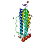
NMR representativeSelection criteria: fewest violations
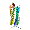
NMR ensembleConformer selection criteria: structures with the least restraint violations
Conformers calculated total number: 100 / Conformers submitted total number: 17 / Representative conformer: 1

+
About Yorodumi

-
News

-
Feb 9, 2022. New format data for meta-information of EMDB entries

New format data for meta-information of EMDB entries

  • Version 3 of the EMDB header file is now the official format.
  • The previous official version 1.9 will be removed from the archive.

Related info.:EMDB header

External links:wwPDB to switch to version 3 of the EMDB data model

-
Aug 12, 2020. Covid-19 info

Covid-19 info

URL: https://pdbjlvh1.pdbj.org/emnavi/covid19.php

New page: Covid-19 featured information page in EM Navigator.

Related info.:Covid-19 info / Mar 5, 2020. Novel coronavirus structure data

+
Mar 5, 2020. Novel coronavirus structure data

Novel coronavirus structure data

Related info.:Yorodumi Speices / Aug 12, 2020. Covid-19 info

External links:COVID-19 featured content - PDBj / Molecule of the Month (242):Coronavirus Proteases

+
Jan 31, 2019. EMDB accession codes are about to change! (news from PDBe EMDB page)

EMDB accession codes are about to change! (news from PDBe EMDB page)

  • The allocation of 4 digits for EMDB accession codes will soon come to an end. Whilst these codes will remain in use, new EMDB accession codes will include an additional digit and will expand incrementally as the available range of codes is exhausted. The current 4-digit format prefixed with “EMD-” (i.e. EMD-XXXX) will advance to a 5-digit format (i.e. EMD-XXXXX), and so on. It is currently estimated that the 4-digit codes will be depleted around Spring 2019, at which point the 5-digit format will come into force.
  • The EM Navigator/Yorodumi systems omit the EMD- prefix.

Related info.:Q: What is EMD? / ID/Accession-code notation in Yorodumi/EM Navigator

External links:EMDB Accession Codes are Changing Soon! / Contact to PDBj

+
Jul 12, 2017. Major update of PDB

Major update of PDB

  • wwPDB released updated PDB data conforming to the new PDBx/mmCIF dictionary.
  • This is a major update changing the version number from 4 to 5, and with Remediation, in which all the entries are updated.
  • In this update, many items about electron microscopy experimental information are reorganized (e.g. em_software).
  • Now, EM Navigator and Yorodumi are based on the updated data.

External links:wwPDB Remediation / Enriched Model Files Conforming to OneDep Data Standards Now Available in the PDB FTP Archive

-
Yorodumi

Thousand views of thousand structures

  • Yorodumi is a browser for structure data from EMDB, PDB, SASBDB, etc.
  • This page is also the successor to EM Navigator detail page, and also detail information page/front-end page for Omokage search.
  • The word "yorodu" (or yorozu) is an old Japanese word meaning "ten thousand". "mi" (miru) is to see.

Related info.:EMDB / PDB / SASBDB / Comparison of 3 databanks / Yorodumi Search / Aug 31, 2016. New EM Navigator & Yorodumi / Yorodumi Papers / Jmol/JSmol / Function and homology information / Changes in new EM Navigator and Yorodumi

Read more