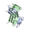
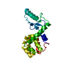
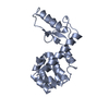
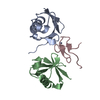
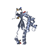
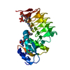
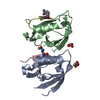
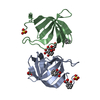
Text: STRUCTURE DETERMINED BY TRIPLE RESONANCE NMR SPECTROSCOPY. NOESY ASSIGNMENTS MADE WITH ITERATIVE METHOD USING CHEMICAL SHIFTS (TALOS) FOR DIHEDRAL ANGLE INFERENCE, AND DYANA FOR SIMULATED ...Text: STRUCTURE DETERMINED BY TRIPLE RESONANCE NMR SPECTROSCOPY. NOESY ASSIGNMENTS MADE WITH ITERATIVE METHOD USING CHEMICAL SHIFTS (TALOS) FOR DIHEDRAL ANGLE INFERENCE, AND DYANA FOR SIMULATED ANNEALING MD LOWEST TARGET FUNCTION SELECTED. CONVERGED STRUCTURES ARE FURTHER REFINED USING NIH-XPLOR FOLLOWED BY CNS IN EXPLICIT WATER SHELL (NIELGES PROTOCOL WITH PARAM19). ASSIGNMENT STATS (EXCLUDING N-TERM TAG): BACKBONE 99.19%, SIDECHAIN 94.31%, AROMATIC (SC) 100%, VL METHYL STEREOSPECIFIC 100%, UNAMBIGUOUS SIDECHAIN NH2 100%. STRUCTURE BASED ON 1252 NOE, 44 H-BOND, 84 DIHEDRAL. 100 STRUCTURES CALCULATED 20 LOWEST ENERGY SUBMITTED. MAX NOE VIOLATION 0.30 A (1 MODEL), MAX DIHEDRAL VIOLATION 4.0 DEG. 5 CLOSE CONTACTS TOTAL PER 20 MODELS. STRUCTURE QUALITY FACTOR PSVS 1.3: ORDERED RESIDUES RANGES - ALPHA HELIX (40-47, 78-88), B-STRAND (53-61, 66-75, 27-32, 99-101) [S(PHI)+S(PSI)] > 1.8. RMSD 0.4 BB, 1.0 ALL HEAVY ATOMS. RAMACHANDRAN: 85.4% MOST FAV, 12.4% ADDTL ALLOW, 2.3 GENEROUSLY ALLOW, 0.0% DISALLOW. PROCHECK (PSI-PHI): -0.46/-1.49 (RAW/Z), PROCHECK (ALL): -0.24/-1.42 (RAW/Z), MOLPROBITY CLASH: 15.06/-1.06 (RAW/Z). RPF SCORES ALL ASSIGNED RESIDUES (FIT OF NOESY PEAKLISTS TO STRUCTURE): RECALL: 0.959, PRECISION: 0.935, F-MEASURE: 0.947, DP-SCORE: 0.775.
-
Sample preparation
Details
Solution-ID
Contents
Solvent system
1
1.3 mM [U-100% 13C; U-100% 15N] protein, 0.02 % sodium azide, 20 mM MES, 100 mM sodium chloride, 5 mM Calcium Chloride, 10 mM DTT, 95% H2O/5% D2O
95% H2O/5% D2O
2
1.1 mM [U-5% 13C; U-100% 15N] protein, 0.02 % sodium azide, 20 mM MES, 100 mM sodium chloride, 5 mM Calcium Chloride, 10 mM DTT, 95% H2O/5% D2O
In the structure databanks used in Yorodumi, some data are registered as the other names, "COVID-19 virus" and "2019-nCoV". Here are the details of the virus and the list of structure data.
Jan 31, 2019. EMDB accession codes are about to change! (news from PDBe EMDB page)
EMDB accession codes are about to change! (news from PDBe EMDB page)
The allocation of 4 digits for EMDB accession codes will soon come to an end. Whilst these codes will remain in use, new EMDB accession codes will include an additional digit and will expand incrementally as the available range of codes is exhausted. The current 4-digit format prefixed with “EMD-” (i.e. EMD-XXXX) will advance to a 5-digit format (i.e. EMD-XXXXX), and so on. It is currently estimated that the 4-digit codes will be depleted around Spring 2019, at which point the 5-digit format will come into force.
The EM Navigator/Yorodumi systems omit the EMD- prefix.
Related info.:Q: What is EMD? / ID/Accession-code notation in Yorodumi/EM Navigator
Yorodumi is a browser for structure data from EMDB, PDB, SASBDB, etc.
This page is also the successor to EM Navigator detail page, and also detail information page/front-end page for Omokage search.
The word "yorodu" (or yorozu) is an old Japanese word meaning "ten thousand". "mi" (miru) is to see.
Related info.:EMDB / PDB / SASBDB / Comparison of 3 databanks / Yorodumi Search / Aug 31, 2016. New EM Navigator & Yorodumi / Yorodumi Papers / Jmol/JSmol / Function and homology information / Changes in new EM Navigator and Yorodumi
 Movie
Movie Controller

 Yorodumi
 Open data
 Basic information
 Components
 Keywords
 Function and homology information
 Homo sapiens (human)
 Authors
 Citation
 Structure visualization
 Downloads & links
 Other downloads
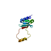

 PDBj
PDBj


 Assembly
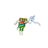

 HSQC
HSQC Sample preparation
 Processing