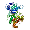
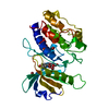
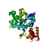
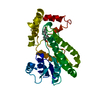
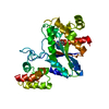
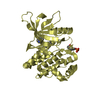
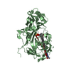
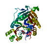
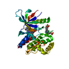
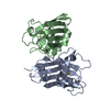
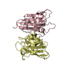
[English] 日本語
Yorodumi
- PDB-2jlp: Crystal structure of human extracellular copper-zinc superoxide d... -

+
Open data


ID or keywords:

Loading...

-
Basic information

Entry
Database: PDB / ID: 2jlp
TitleCrystal structure of human extracellular copper-zinc superoxide dismutase.
ComponentsEXTRACELLULAR SUPEROXIDE DISMUTASE (CU-ZN)
KeywordsOXIDOREDUCTASE / GLYCOLISATION / HEPARIN BINDING / HEPARIN-BINDING / OXIDATIVE STRESS / ANTIOXIDANT / GLYCOPROTEIN / METAL-BINDING / CU-ZN / SECRETED / GLYCATION
Function / homology
Function and homology information


response to copper ion / superoxide dismutase / Detoxification of Reactive Oxygen Species / superoxide dismutase activity / removal of superoxide radicals / Golgi lumen / heparin binding / cellular response to oxidative stress / collagen-containing extracellular matrix / response to hypoxia ...response to copper ion / superoxide dismutase / Detoxification of Reactive Oxygen Species / superoxide dismutase activity / removal of superoxide radicals / Golgi lumen / heparin binding / cellular response to oxidative stress / collagen-containing extracellular matrix / response to hypoxia / copper ion binding / extracellular space / extracellular exosome / extracellular region / nucleus
Similarity search - Function
Superoxide dismutase, copper/zinc binding domain / Copper/Zinc superoxide dismutase signature 1. / Superoxide dismutase, copper/zinc, binding site / Copper/Zinc superoxide dismutase signature 2. / Superoxide dismutase, copper/zinc binding domain / Copper/zinc superoxide dismutase (SODC) / Superoxide dismutase (Cu/Zn) / superoxide dismutase copper chaperone / Superoxide dismutase-like, copper/zinc binding domain superfamily / Immunoglobulin-like / Sandwich / Mainly Beta
Similarity search - Domain/homology
COPPER (II) ION / THIOCYANATE ION / Extracellular superoxide dismutase [Cu-Zn]
Similarity search - Component
Biological speciesHOMO SAPIENS (human)
MethodX-RAY DIFFRACTION / SYNCHROTRON / MOLECULAR REPLACEMENT / Resolution: 1.7 Å
AuthorsAntonyuk, S.V. / Strange, R.W. / Marklund, S.L. / Hasnain, S.S.
CitationJournal: J.Mol.Biol. / Year: 2009
Title: The Structure of Human Extracellular Copper-Zinc Superoxide Dismutase at 1.7 A Resolution: Insights Into Heparin and Collagen Binding.
Authors: Antonyuk, S.V. / Strange, R.W. / Marklund, S.L. / Hasnain, S.S.
History
DepositionSep 14, 2008Deposition site: PDBE / Processing site: PDBE
Revision 1.0Mar 17, 2009Provider: repository / Type: Initial release
Revision 1.1Jul 13, 2011Group: Advisory / Version format compliance
Revision 1.2Apr 3, 2019Group: Data collection / Other / Source and taxonomy
Category: entity_src_gen / pdbx_database_proc / pdbx_database_status
Item: _entity_src_gen.pdbx_host_org_cell_line / _pdbx_database_status.recvd_author_approval
Revision 1.3Mar 4, 2020Group: Derived calculations / Other
Category: pdbx_database_status / pdbx_struct_assembly ...pdbx_database_status / pdbx_struct_assembly / pdbx_struct_assembly_gen / pdbx_struct_assembly_prop
Item: _pdbx_database_status.status_code_sf / _pdbx_struct_assembly_prop.biol_id / _pdbx_struct_assembly_prop.value
Revision 1.4Dec 13, 2023Group: Data collection / Database references ...Data collection / Database references / Derived calculations / Refinement description
Category: chem_comp_atom / chem_comp_bond ...chem_comp_atom / chem_comp_bond / database_2 / pdbx_initial_refinement_model / pdbx_struct_conn_angle / struct_conn / struct_site
Item: _database_2.pdbx_DOI / _database_2.pdbx_database_accession ..._database_2.pdbx_DOI / _database_2.pdbx_database_accession / _pdbx_struct_conn_angle.ptnr1_auth_comp_id / _pdbx_struct_conn_angle.ptnr1_auth_seq_id / _pdbx_struct_conn_angle.ptnr1_label_asym_id / _pdbx_struct_conn_angle.ptnr1_label_atom_id / _pdbx_struct_conn_angle.ptnr1_label_comp_id / _pdbx_struct_conn_angle.ptnr1_label_seq_id / _pdbx_struct_conn_angle.ptnr2_label_alt_id / _pdbx_struct_conn_angle.ptnr3_auth_comp_id / _pdbx_struct_conn_angle.ptnr3_auth_seq_id / _pdbx_struct_conn_angle.ptnr3_label_asym_id / _pdbx_struct_conn_angle.ptnr3_label_atom_id / _pdbx_struct_conn_angle.ptnr3_label_comp_id / _pdbx_struct_conn_angle.ptnr3_label_seq_id / _pdbx_struct_conn_angle.value / _struct_conn.pdbx_dist_value / _struct_conn.pdbx_ptnr1_label_alt_id / _struct_conn.pdbx_ptnr2_label_alt_id / _struct_conn.ptnr1_auth_comp_id / _struct_conn.ptnr1_auth_seq_id / _struct_conn.ptnr1_label_asym_id / _struct_conn.ptnr1_label_atom_id / _struct_conn.ptnr1_label_comp_id / _struct_conn.ptnr1_label_seq_id / _struct_conn.ptnr2_auth_comp_id / _struct_conn.ptnr2_auth_seq_id / _struct_conn.ptnr2_label_asym_id / _struct_conn.ptnr2_label_atom_id / _struct_conn.ptnr2_label_comp_id / _struct_conn.ptnr2_label_seq_id / _struct_site.pdbx_auth_asym_id / _struct_site.pdbx_auth_comp_id / _struct_site.pdbx_auth_seq_id
Remark 700 SHEET DETERMINATION METHOD: DSSP THE SHEETS PRESENTED AS "CA" IN EACH CHAIN ON SHEET RECORDS BELOW ... SHEET DETERMINATION METHOD: DSSP THE SHEETS PRESENTED AS "CA" IN EACH CHAIN ON SHEET RECORDS BELOW IS ACTUALLY AN 9-STRANDED BARREL THIS IS REPRESENTED BY A 10-STRANDED SHEET IN WHICH THE FIRST AND LAST STRANDS ARE IDENTICAL. THE SHEET STRUCTURE OF THIS MOLECULE IS BIFURCATED. IN ORDER TO REPRESENT THIS FEATURE IN THE SHEET RECORDS BELOW, TWO SHEETS ARE DEFINED.

-
Structure visualization

Structure viewerMolecule:
MolmilJmol/JSmol

Downloads & links

-
Assembly

Deposited unit
A: EXTRACELLULAR SUPEROXIDE DISMUTASE (CU-ZN)
B: EXTRACELLULAR SUPEROXIDE DISMUTASE (CU-ZN)
C: EXTRACELLULAR SUPEROXIDE DISMUTASE (CU-ZN)
D: EXTRACELLULAR SUPEROXIDE DISMUTASE (CU-ZN)
hetero molecules


Theoretical massNumber of molelcules
Total (without water)97,41914
Polymers96,7874
Non-polymers63210
Water16,015889
1
A: EXTRACELLULAR SUPEROXIDE DISMUTASE (CU-ZN)
B: EXTRACELLULAR SUPEROXIDE DISMUTASE (CU-ZN)
hetero molecules


Theoretical massNumber of molelcules
Total (without water)48,7107
Polymers48,3942
Non-polymers3165
Water362
TypeNameSymmetry operationNumber
identity operation1_555x,y,z1
Buried area1560 Å2
ΔGint-17 kcal/mol
Surface area15040 Å2
MethodPISA
2
C: EXTRACELLULAR SUPEROXIDE DISMUTASE (CU-ZN)
D: EXTRACELLULAR SUPEROXIDE DISMUTASE (CU-ZN)
hetero molecules


Theoretical massNumber of molelcules
Total (without water)48,7107
Polymers48,3942
Non-polymers3165
Water362
TypeNameSymmetry operationNumber
identity operation1_555x,y,z1
Buried area1590 Å2
ΔGint-17 kcal/mol
Surface area15310 Å2
MethodPISA
Unit cell
Length a, b, c (Å)56.738, 93.588, 75.600
Angle α, β, γ (deg.)90.00, 106.23, 90.00
Int Tables number4
Space group name H-MP1211

-
Components

#1: Protein
EXTRACELLULAR SUPEROXIDE DISMUTASE (CU-ZN) / EXTRACELLULAR COPPER-ZINC SUPEROXIDE DISMUTASE / EC-SOD


Mass: 24196.838 Da / Num. of mol.: 4
Source method: isolated from a genetically manipulated source
Source: (gene. exp.) HOMO SAPIENS (human) / Cell line (production host): CHO / Production host: 
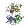
CRICETULUS GRISEUS (Chinese hamster) / Tissue (production host): OVARY / References: UniProt: P08294, superoxide dismutase
#2: Chemical
ChemComp-CU / COPPER (II) ION


Mass: 63.546 Da / Num. of mol.: 4 / Source method: obtained synthetically / Formula: Cu
#3: Chemical
ChemComp-ZN / ZINC ION


Mass: 65.409 Da / Num. of mol.: 4 / Source method: obtained synthetically / Formula: Zn
#4: Chemical ChemComp-SCN / THIOCYANATE ION


Mass: 58.082 Da / Num. of mol.: 2 / Source method: obtained synthetically / Formula: CNS
#5: Water ChemComp-HOH / water


Mass: 18.015 Da / Num. of mol.: 889 / Source method: isolated from a natural source / Formula: H2O
Sequence detailsELECTRON DENSITY AND WATER NETWORK IS IN AGREEMENT WITH THR IN POSITION 40

-
Experimental details

-
Experiment

ExperimentMethod: X-RAY DIFFRACTION / Number of used crystals: 1

-
Sample preparation

CrystalDensity Matthews: 1.93 Å3/Da / Density % sol: 36.2 % / Description: NONE
Crystal growpH: 7.5 / Details: PEG3350, THIOCYANATE, BIS-TRIS PROPANE PH 7.5

-
Data collection

DiffractionMean temperature: 100 K
Diffraction sourceSource: SYNCHROTRON / Site: SRS / Beamline: PX10.1 / Wavelength: 0.98
DetectorType: MARRESEARCH / Detector: CCD / Date: Sep 22, 2007 / Details: MIRRORS
RadiationMonochromator: SI111 / Protocol: SINGLE WAVELENGTH / Monochromatic (M) / Laue (L): M / Scattering type: x-ray
Radiation wavelengthWavelength: 0.98 Å / Relative weight: 1
ReflectionResolution: 1.7→28.2 Å / Num. obs: 81056 / % possible obs: 97.3 % / Redundancy: 3.7 % / Biso Wilson estimate: 20.8 Å2 / Rmerge(I) obs: 0.06 / Net I/σ(I): 16
Reflection shellResolution: 1.7→1.76 Å / Redundancy: 2.8 % / Rmerge(I) obs: 0.3 / Mean I/σ(I) obs: 3 / % possible all: 80

-
Processing

Software
NameVersionClassification
REFMAC5.3.0037refinement
HKL-2000data reduction
HKL-2000data scaling
MOLREPphasing
RefinementMethod to determine structure: MOLECULAR REPLACEMENT
Starting model: PDB ENTRY 2C9V
Resolution: 1.7→28.2 Å / Cor.coef. Fo:Fc: 0.972 / Cor.coef. Fo:Fc free: 0.96 / SU B: 3.546 / SU ML: 0.061 / TLS residual ADP flag: LIKELY RESIDUAL / Cross valid method: THROUGHOUT / ESU R: 0.088 / ESU R Free: 0.09 / Stereochemistry target values: MAXIMUM LIKELIHOOD / Details: HYDROGENS HAVE BEEN ADDED IN THE RIDING POSITIONS.
RfactorNum. reflection% reflectionSelection details
Rfree0.185 4057 5 %RANDOM
Rwork0.15 ---
obs0.152 76936 97.2 %-
Solvent computationIon probe radii: 0.8 Å / Shrinkage radii: 0.8 Å / VDW probe radii: 1.4 Å / Solvent model: MASK
Displacement parametersBiso mean: 24.02 Å2
Baniso -1Baniso -2Baniso -3
1--1.24 Å20 Å2-0.56 Å2
2--1.29 Å20 Å2
3----0.37 Å2
Refinement stepCycle: LAST / Resolution: 1.7→28.2 Å
ProteinNucleic acidLigandSolventTotal
Num. atoms4954 0 14 889 5857
Refine LS restraints
Refine-IDTypeDev idealDev ideal targetNumber
X-RAY DIFFRACTIONr_bond_refined_d0.0180.0215245
X-RAY DIFFRACTIONr_bond_other_d0.0020.023585
X-RAY DIFFRACTIONr_angle_refined_deg1.711.9357162
X-RAY DIFFRACTIONr_angle_other_deg0.9963.0038632
X-RAY DIFFRACTIONr_dihedral_angle_1_deg6.8375706
X-RAY DIFFRACTIONr_dihedral_angle_2_deg34.622.549255
X-RAY DIFFRACTIONr_dihedral_angle_3_deg11.71115734
X-RAY DIFFRACTIONr_dihedral_angle_4_deg16.2871557
X-RAY DIFFRACTIONr_chiral_restr0.1140.2752
X-RAY DIFFRACTIONr_gen_planes_refined0.0080.026169
X-RAY DIFFRACTIONr_gen_planes_other0.0020.021144
X-RAY DIFFRACTIONr_nbd_refined0.2620.21096
X-RAY DIFFRACTIONr_nbd_other0.2280.24127
X-RAY DIFFRACTIONr_nbtor_refined0.1690.22502
X-RAY DIFFRACTIONr_nbtor_other0.0860.22831
X-RAY DIFFRACTIONr_xyhbond_nbd_refined0.1980.2615
X-RAY DIFFRACTIONr_xyhbond_nbd_other
X-RAY DIFFRACTIONr_metal_ion_refined
X-RAY DIFFRACTIONr_metal_ion_other
X-RAY DIFFRACTIONr_symmetry_vdw_refined0.1340.220
X-RAY DIFFRACTIONr_symmetry_vdw_other0.3120.281
X-RAY DIFFRACTIONr_symmetry_hbond_refined0.1680.234
X-RAY DIFFRACTIONr_symmetry_hbond_other
X-RAY DIFFRACTIONr_symmetry_metal_ion_refined
X-RAY DIFFRACTIONr_symmetry_metal_ion_other
X-RAY DIFFRACTIONr_mcbond_it1.011.53384
X-RAY DIFFRACTIONr_mcbond_other
X-RAY DIFFRACTIONr_mcangle_it1.66725376
X-RAY DIFFRACTIONr_mcangle_other
X-RAY DIFFRACTIONr_scbond_it2.47531861
X-RAY DIFFRACTIONr_scbond_other
X-RAY DIFFRACTIONr_scangle_it3.8344.51768
X-RAY DIFFRACTIONr_scangle_other
X-RAY DIFFRACTIONr_long_range_B_refined
X-RAY DIFFRACTIONr_long_range_B_other
X-RAY DIFFRACTIONr_rigid_bond_restr
X-RAY DIFFRACTIONr_sphericity_free
X-RAY DIFFRACTIONr_sphericity_bonded
LS refinement shellResolution: 1.7→1.74 Å / Total num. of bins used: 20
RfactorNum. reflection% reflection
Rfree0.283 221 -
Rwork0.233 4520 -
obs--76.94 %
Refinement TLS params.

Method: refined / Refine-ID: X-RAY DIFFRACTION

IDL112)L122)L132)L222)L232)L332)S11 (Å °)S12 (Å °)S13 (Å °)S21 (Å °)S22 (Å °)S23 (Å °)S31 (Å °)S32 (Å °)S33 (Å °)T112)T122)T132)T222)T232)T332)Origin x (Å)Origin y (Å)Origin z (Å)
11.6876-0.3396-0.04171.3508-0.06391.41290.01540.16160.0776-0.02470.02550.1021-0.05-0.086-0.0408-0.17370.00680.001-0.15940.043-0.159521.985-0.01116.451
21.96170.39080.58321.84080.71561.79520.02650.3487-0.162-0.07950.1249-0.3091-0.00630.1864-0.1514-0.15540.0040.0168-0.0997-0.0628-0.100442.348-39.68212.747
32.8471-0.0944-0.21962.13830.8181.71260.016-0.0905-0.15890.3342-0.10960.16720.2323-0.16490.0936-0.1183-0.03180.034-0.1726-0.0064-0.154420.728-37.19927.711
43.4376-0.63830.34651.8127-0.28421.3166-0.1081-0.2170.01480.2760.0031-0.188-0.1060.08950.1051-0.1356-0.0006-0.0359-0.18210.0239-0.129644.066-5.35630.422
Refinement TLS group
IDRefine-IDRefine TLS-IDAuth asym-IDAuth seq-ID
1X-RAY DIFFRACTION1A39 - 204
2X-RAY DIFFRACTION2D39 - 204
3X-RAY DIFFRACTION3C39 - 204
4X-RAY DIFFRACTION4B39 - 204

+
About Yorodumi

-
News

-
Feb 9, 2022. New format data for meta-information of EMDB entries

New format data for meta-information of EMDB entries

  • Version 3 of the EMDB header file is now the official format.
  • The previous official version 1.9 will be removed from the archive.

Related info.:EMDB header

External links:wwPDB to switch to version 3 of the EMDB data model

-
Aug 12, 2020. Covid-19 info

Covid-19 info

URL: https://pdbjlvh1.pdbj.org/emnavi/covid19.php

New page: Covid-19 featured information page in EM Navigator.

Related info.:Covid-19 info / Mar 5, 2020. Novel coronavirus structure data

+
Mar 5, 2020. Novel coronavirus structure data

Novel coronavirus structure data

Related info.:Yorodumi Speices / Aug 12, 2020. Covid-19 info

External links:COVID-19 featured content - PDBj / Molecule of the Month (242):Coronavirus Proteases

+
Jan 31, 2019. EMDB accession codes are about to change! (news from PDBe EMDB page)

EMDB accession codes are about to change! (news from PDBe EMDB page)

  • The allocation of 4 digits for EMDB accession codes will soon come to an end. Whilst these codes will remain in use, new EMDB accession codes will include an additional digit and will expand incrementally as the available range of codes is exhausted. The current 4-digit format prefixed with “EMD-” (i.e. EMD-XXXX) will advance to a 5-digit format (i.e. EMD-XXXXX), and so on. It is currently estimated that the 4-digit codes will be depleted around Spring 2019, at which point the 5-digit format will come into force.
  • The EM Navigator/Yorodumi systems omit the EMD- prefix.

Related info.:Q: What is EMD? / ID/Accession-code notation in Yorodumi/EM Navigator

External links:EMDB Accession Codes are Changing Soon! / Contact to PDBj

+
Jul 12, 2017. Major update of PDB

Major update of PDB

  • wwPDB released updated PDB data conforming to the new PDBx/mmCIF dictionary.
  • This is a major update changing the version number from 4 to 5, and with Remediation, in which all the entries are updated.
  • In this update, many items about electron microscopy experimental information are reorganized (e.g. em_software).
  • Now, EM Navigator and Yorodumi are based on the updated data.

External links:wwPDB Remediation / Enriched Model Files Conforming to OneDep Data Standards Now Available in the PDB FTP Archive

-
Yorodumi

Thousand views of thousand structures

  • Yorodumi is a browser for structure data from EMDB, PDB, SASBDB, etc.
  • This page is also the successor to EM Navigator detail page, and also detail information page/front-end page for Omokage search.
  • The word "yorodu" (or yorozu) is an old Japanese word meaning "ten thousand". "mi" (miru) is to see.

Related info.:EMDB / PDB / SASBDB / Comparison of 3 databanks / Yorodumi Search / Aug 31, 2016. New EM Navigator & Yorodumi / Yorodumi Papers / Jmol/JSmol / Function and homology information / Changes in new EM Navigator and Yorodumi

Read more