 Movie
Movie Controller

[English] 日本語
 Yorodumi
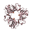
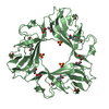
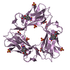
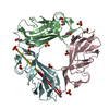
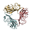
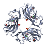
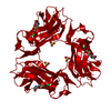
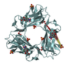
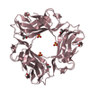
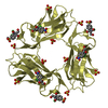
- PDB-2jkn: DraE Adhesin in complex with Chloramphenicol Succinate (trigonal form) -
+ Open data

- Basic information
| Entry | Database: PDB / ID: 2jkn | ||||||
|---|---|---|---|---|---|---|---|
| Title | DraE Adhesin in complex with Chloramphenicol Succinate (trigonal form) | ||||||
 Components Components | DR HEMAGGLUTININ STRUCTURAL SUBUNIT | ||||||
 Keywords Keywords | CELL ADHESION / UPEC / DRAE / DAEC / ADHESIN / FIMBRIUM / HAEMAGGLUTININ / CELL PROJECTION / FIMBRIAL ADHESIN / CHLORAMPHENICOL SUCCINATE | ||||||
| Function / homology |  Function and homology information Function and homology information | ||||||
| Biological species |  | ||||||
| Method |  X-RAY DIFFRACTION / MOLECULAR REPLACEMENT / Resolution: 1.9 Å X-RAY DIFFRACTION / MOLECULAR REPLACEMENT / Resolution: 1.9 Å | ||||||
 Authors Authors | Pettigrew, D.M. / Roversi, P. / Davies, S.G. / Russell, A.J. / Lea, S.M. | ||||||
 Citation Citation | Journal: Acta Crystallogr. D Biol. Crystallogr. / Year: 2009 Title: A structural study of the interaction between the Dr haemagglutinin DraE and derivatives of chloramphenicol. Authors: Pettigrew, D.M. / Roversi, P. / Davies, S.G. / Russell, A.J. / Lea, S.M. | ||||||
| History |
| ||||||
| Remark 700 | SHEET THE SHEET STRUCTURE OF THIS MOLECULE IS BIFURCATED. IN ORDER TO REPRESENT THIS FEATURE IN ... SHEET THE SHEET STRUCTURE OF THIS MOLECULE IS BIFURCATED. IN ORDER TO REPRESENT THIS FEATURE IN THE SHEET RECORDS BELOW, TWO SHEETS ARE DEFINED. |
- Structure visualization
| Structure viewer | Molecule: MolmilJmol/JSmol |
|---|
- Downloads & links
-Download
| PDBx/mmCIF format | 2jkn.cif.gz | 191.3 KB | Display | PDBx/mmCIF format |
|---|---|---|---|---|
| PDB format | pdb2jkn.ent.gz | 155.6 KB | Display | PDB format |
| PDBx/mmJSON format | 2jkn.json.gz | Tree view | PDBx/mmJSON format | |
| Others |  Other downloads Other downloads |
-Validation report
| Arichive directory | https://data.pdbj.org/pub/pdb/validation_reports/jk/2jknftp://data.pdbj.org/pub/pdb/validation_reports/jk/2jkn | HTTPS FTP |
|---|
-Related structure data
| Related structure data |  2jkjC  2jklC  2w5pC 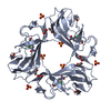 1usqS S: Starting model for refinement C: citing same article ( |
|---|---|
| Similar structure data |


-Links
 PDBj
PDBj- Assembly
| Deposited unit | 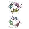
| ||||||||||||||||||||||||
|---|---|---|---|---|---|---|---|---|---|---|---|---|---|---|---|---|---|---|---|---|---|---|---|---|---|
| 1 | 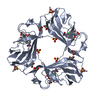
| ||||||||||||||||||||||||
| 2 | 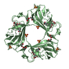
| ||||||||||||||||||||||||
| 3 | 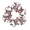
| ||||||||||||||||||||||||
| 4 | 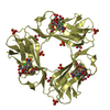
| ||||||||||||||||||||||||
| 5 | 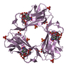
| ||||||||||||||||||||||||
| 6 | 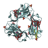
| ||||||||||||||||||||||||
| Unit cell |
| ||||||||||||||||||||||||
| Noncrystallographic symmetry (NCS) | NCS oper:
|
-Components
| #1: Protein | Mass: 16169.844 Da / Num. of mol.: 6 / Fragment: ADHESIN SUBUNIT, RESIDUES 23-160 Source method: isolated from a genetically manipulated source Source: (gene. exp.) #2: Chemical | ChemComp-SO4 /   Mass: 96.063 Da / Num. of mol.: 12 / Source method: obtained synthetically / Formula: SO4 Mass: 96.063 Da / Num. of mol.: 12 / Source method: obtained synthetically / Formula: SO4#3: Chemical | ChemComp-CL8 / 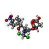  Mass: 423.202 Da / Num. of mol.: 6 / Source method: obtained synthetically / Formula: C15H16Cl2N2O8 Mass: 423.202 Da / Num. of mol.: 6 / Source method: obtained synthetically / Formula: C15H16Cl2N2O8#4: Chemical | ChemComp-EDO /   Mass: 62.068 Da / Num. of mol.: 12 / Source method: obtained synthetically / Formula: C2H6O2 Mass: 62.068 Da / Num. of mol.: 12 / Source method: obtained synthetically / Formula: C2H6O2#5: Water | ChemComp-HOH / |  Mass: 18.015 Da / Num. of mol.: 702 / Source method: isolated from a natural source / Formula: H2O Mass: 18.015 Da / Num. of mol.: 702 / Source method: isolated from a natural source / Formula: H2OHas protein modification | Y | |
|---|
-Experimental details
-Experiment
| Experiment | Method: X-RAY DIFFRACTION / Number of used crystals: 1 |
|---|
- Sample preparation
| Crystal | Density Matthews: 2.56 Å3/Da / Density % sol: 52 % / Description: NONE |
|---|---|
| Crystal grow | Details: 2M AMMONIUM SULPHATE, 0.1-20 MM CHLORAMPHENICOL SUCCINATE, 0.1 M HEPES PH 7.0 |
-Data collection
| Diffraction | Mean temperature: 100 K |
|---|---|
| Diffraction source | Source: ROTATING ANODE / Type: RIGAKU FR-D / Wavelength: 1.5418 |
| Detector | Type: MARRESEARCH / Detector: CCD / Date: Oct 26, 2004 |
| Radiation | Protocol: SINGLE WAVELENGTH / Monochromatic (M) / Laue (L): M / Scattering type: x-ray |
| Radiation wavelength | Wavelength: 1.5418 Å / Relative weight: 1 |
| Reflection | Resolution: 1.9→30 Å / Num. obs: 60758 / % possible obs: 98.2 % / Observed criterion σ(I): 0 / Redundancy: 3.3 % / Rmerge(I) obs: 0.07 / Net I/σ(I): 4.6 |
| Reflection shell | Resolution: 1.9→2 Å / Redundancy: 3.3 % / Rmerge(I) obs: 0.21 / Mean I/σ(I) obs: 2.1 / % possible all: 97.7 |
- Processing
| Software |
| ||||||||||||||||||||
|---|---|---|---|---|---|---|---|---|---|---|---|---|---|---|---|---|---|---|---|---|---|
| Refinement | Method to determine structure: MOLECULAR REPLACEMENT Starting model: PDB ENTRY 1USQ Resolution: 1.9→30 Å / Cross valid method: THROUGHOUT / σ(F): 0
| ||||||||||||||||||||
| Refinement step | Cycle: LAST / Resolution: 1.9→30 Å
|
