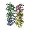
 Movie
Movie Controller

[English] 日本語
 Yorodumi
- PDB-2jjm: Crystal Structure of a family GT4 glycosyltransferase from Bacill... -
+ Open data

- Basic information
| Entry | Database: PDB / ID: 2jjm | ||||||
|---|---|---|---|---|---|---|---|
| Title | Crystal Structure of a family GT4 glycosyltransferase from Bacillus anthracis ORF BA1558. | ||||||
 Components Components | GLYCOSYL TRANSFERASE, GROUP 1 FAMILY PROTEIN | ||||||
 Keywords Keywords | TRANSFERASE / GLYCOSYL TRANSFER / GLYCOSYLTRANSFERASE / ANTHRAX / NUCLEOTIDE / CARBOHYDRATE | ||||||
| Function / homology |  Function and homology information Function and homology informationbacillithiol biosynthetic process / Transferases; Glycosyltransferases; Hexosyltransferases / glycosyltransferase activity / nucleotide binding Similarity search - Function | ||||||
| Biological species |  | ||||||
| Method |  X-RAY DIFFRACTION / SYNCHROTRON / SAD / Resolution: 3.1 Å X-RAY DIFFRACTION / SYNCHROTRON / SAD / Resolution: 3.1 Å | ||||||
 Authors Authors | Ruane, K.M. / Davies, G.J. / Martinez-Fleites, C. | ||||||
 Citation Citation | Journal: Proteins / Year: 2008 Title: Crystal Structure of a Family Gt4 Glycosyltransferase from Bacillus Anthracis Orf Ba1558. Authors: Ruane, K.M. / Davies, G.J. / Martinez-Fleites, C. | ||||||
| History |
|
- Structure visualization
| Structure viewer | Molecule: MolmilJmol/JSmol |
|---|
- Downloads & links
-Download
| PDBx/mmCIF format | 2jjm.cif.gz | 806.2 KB | Display | PDBx/mmCIF format |
|---|---|---|---|---|
| PDB format | pdb2jjm.ent.gz | 677.5 KB | Display | PDB format |
| PDBx/mmJSON format | 2jjm.json.gz | Tree view | PDBx/mmJSON format | |
| Others |  Other downloads Other downloads |
-Validation report
| Summary document | 2jjm_validation.pdf.gz | 566.1 KB | Display | wwPDB validaton report |
|---|---|---|---|---|
| Full document | 2jjm_full_validation.pdf.gz | 777.5 KB | Display | |
| Data in XML | 2jjm_validation.xml.gz | 172.9 KB | Display | |
| Data in CIF | 2jjm_validation.cif.gz | 223.7 KB | Display | |
| Arichive directory | https://data.pdbj.org/pub/pdb/validation_reports/jj/2jjmftp://data.pdbj.org/pub/pdb/validation_reports/jj/2jjm | HTTPS FTP |
-Related structure data
| Similar structure data |
|---|


-Links
 PDBj
PDBj
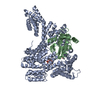
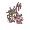
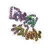
- Assembly
| Deposited unit | 
| ||||||||||||||||||||||||||||||||||||||||||||||||
|---|---|---|---|---|---|---|---|---|---|---|---|---|---|---|---|---|---|---|---|---|---|---|---|---|---|---|---|---|---|---|---|---|---|---|---|---|---|---|---|---|---|---|---|---|---|---|---|---|---|
| 1 | 
| ||||||||||||||||||||||||||||||||||||||||||||||||
| 2 | 
| ||||||||||||||||||||||||||||||||||||||||||||||||
| 3 | 
| ||||||||||||||||||||||||||||||||||||||||||||||||
| Unit cell |
| ||||||||||||||||||||||||||||||||||||||||||||||||
| Noncrystallographic symmetry (NCS) | NCS oper:
|
-Components
| #1: Protein | Mass: 44565.074 Da / Num. of mol.: 12 Source method: isolated from a genetically manipulated source Source: (gene. exp.) |
|---|
-Experimental details
-Experiment
| Experiment | Method: X-RAY DIFFRACTION |
|---|
- Sample preparation
| Crystal | Density Matthews: 3.37 Å3/Da / Density % sol: 63.5 % / Description: NONE |
|---|---|
| Crystal grow | Details: 0.1M HEPES PH 7.5, 0.2M NA2SO4, 14% PEG 3350 |
-Data collection
| Diffraction | Mean temperature: 290 K |
|---|---|
| Diffraction source | Source: SYNCHROTRON / Site: ESRF  / Beamline: ID14-4 / Wavelength: 0.9795 / Beamline: ID14-4 / Wavelength: 0.9795 |
| Detector | Type: ADSC CCD / Detector: CCD |
| Radiation | Protocol: SINGLE WAVELENGTH / Monochromatic (M) / Laue (L): M / Scattering type: x-ray |
| Radiation wavelength | Wavelength: 0.9795 Å / Relative weight: 1 |
| Reflection | Resolution: 3.1→20 Å / Num. obs: 117619 / % possible obs: 98.5 % / Observed criterion σ(I): 2 / Redundancy: 7.5 % / Biso Wilson estimate: 100.415 Å2 / Rmerge(I) obs: 0.11 |
| Reflection shell | Resolution: 3.1→3.27 Å / Redundancy: 7 % / Rmerge(I) obs: 0.87 / Mean I/σ(I) obs: 2.1 / % possible all: 98 |
- Processing
| Software |
| ||||||||||||||||||||||||||||||||||||||||||||||||||||||||||||
|---|---|---|---|---|---|---|---|---|---|---|---|---|---|---|---|---|---|---|---|---|---|---|---|---|---|---|---|---|---|---|---|---|---|---|---|---|---|---|---|---|---|---|---|---|---|---|---|---|---|---|---|---|---|---|---|---|---|---|---|---|---|
| Refinement | Method to determine structure: SAD Starting model: NONE Resolution: 3.1→20 Å / Rfactor Rfree error: 0.003 / Data cutoff high absF: 2903911.94 / Isotropic thermal model: RESTRAINED / Cross valid method: THROUGHOUT / σ(F): 0 / Stereochemistry target values: MAXIMUM LIKELIHOOD Details: BULK SOLVENT MODEL USED DISORDERED REGIONS ARE THE SAME IN EACH MONOMER AND ARE 12- 13 43-46 61- 64 196-198
| ||||||||||||||||||||||||||||||||||||||||||||||||||||||||||||
| Solvent computation | Solvent model: FLAT MODEL / Bsol: 68.263 Å2 / ksol: 0.35 e/Å3 | ||||||||||||||||||||||||||||||||||||||||||||||||||||||||||||
| Displacement parameters | Biso mean: 91.6 Å2
| ||||||||||||||||||||||||||||||||||||||||||||||||||||||||||||
| Refine analyze |
| ||||||||||||||||||||||||||||||||||||||||||||||||||||||||||||
| Refinement step | Cycle: LAST / Resolution: 3.1→20 Å
| ||||||||||||||||||||||||||||||||||||||||||||||||||||||||||||
| Refine LS restraints |
| ||||||||||||||||||||||||||||||||||||||||||||||||||||||||||||
| Refine LS restraints NCS | NCS model details: CONSTR | ||||||||||||||||||||||||||||||||||||||||||||||||||||||||||||
| LS refinement shell | Resolution: 3.1→3.29 Å / Rfactor Rfree error: 0.012 / Total num. of bins used: 6
| ||||||||||||||||||||||||||||||||||||||||||||||||||||||||||||
| Xplor file | Serial no: 1 / Param file: PROTEIN_REP.PARAM / Topol file: PROTEIN.TOP |
