 Movie
Movie Controller

[English] 日本語
 Yorodumi
- PDB-2jba: PhoB response regulator receiver domain constitutively-active dou... -
+ Open data

- Basic information
| Entry | Database: PDB / ID: 2jba | ||||||
|---|---|---|---|---|---|---|---|
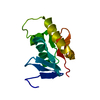
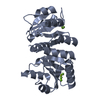
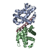
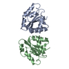
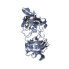
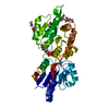
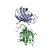
| Title | PhoB response regulator receiver domain constitutively-active double mutant D53A and Y102C. | ||||||
 Components Components | (PHOSPHATE REGULON TRANSCRIPTIONAL REGULATORY PROTEIN PHOB) x 2 | ||||||
 Keywords Keywords | TRANSCRIPTION / TRANSCRIPTION FACTOR / SENSORY TRANSDUCTION / PHOSPHATE REGULATION / TRANSCRIPTION REGULATION / ACTIVATOR / TRANSPORT / DNA-BINDING / CONSTITUTIVELY-ACTIVE MUTANT / TWO-COMPONENT REGULATORY SYSTEM / GENE REGULATION / PHOSPHATE TRANSPORT / ACTIVATION OF THE PHO REGULON / PHOSPHORYLATION / ALPHA/BETA DOUBLY WOUN FOLD | ||||||
| Function / homology |  Function and homology information Function and homology informationbacterial-type RNA polymerase holo enzyme binding / phosphate ion transport / phosphorelay response regulator activity / regulation of DNA-templated transcription initiation / protein-DNA complex / transcription cis-regulatory region binding / regulation of DNA-templated transcription / identical protein binding / cytosol Similarity search - Function | ||||||
| Biological species |  | ||||||
| Method |  X-RAY DIFFRACTION / SYNCHROTRON / MOLECULAR REPLACEMENT / Resolution: 1.45 Å X-RAY DIFFRACTION / SYNCHROTRON / MOLECULAR REPLACEMENT / Resolution: 1.45 Å | ||||||
 Authors Authors | Arribas-Bosacoma, R. / Ferrer-Orta, C. / Kim, S.-K. / Blanco, A.G. / Pereira, P.J.B. / Gomis-Ruth, F.X. / Wanner, B.L. / Coll, M. / Sola, M. | ||||||
 Citation Citation | Journal: J.Mol.Biol. / Year: 2007 Title: The X-Ray Crystal Structures of Two Constitutively Active Mutants of the Escherichia Coli Phob Receiver Domain Give Insights Into Activation. Authors: Arribas-Bosacoma, R. / Kim, S.-K. / Ferrer-Orta, C. / Blanco, A.G. / Pereira, P.J.B. / Gomis-Ruth, F.X. / Wanner, B.L. / Coll, M. / Sola, M. #1: Journal: J.Mol.Biol. / Year: 1999Title: Three-Dimensional Crystal Structure of the Transcription Factor Phob Receiver Domain. Authors: Sola, M. / Gomis-Ruth, F.X. / Serrano, L. / Gonzalez, A. / Coll, M. | ||||||
| History |
|
- Structure visualization
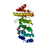
| Structure viewer | Molecule: MolmilJmol/JSmol |
|---|
- Downloads & links
-Download
| PDBx/mmCIF format | 2jba.cif.gz | 72.3 KB | Display | PDBx/mmCIF format |
|---|---|---|---|---|
| PDB format | pdb2jba.ent.gz | 52.7 KB | Display | PDB format |
| PDBx/mmJSON format | 2jba.json.gz | Tree view | PDBx/mmJSON format | |
| Others |  Other downloads Other downloads |
-Validation report
| Arichive directory | https://data.pdbj.org/pub/pdb/validation_reports/jb/2jbaftp://data.pdbj.org/pub/pdb/validation_reports/jb/2jba | HTTPS FTP |
|---|
-Related structure data
| Related structure data | 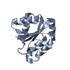 2jb9C  1b00S S: Starting model for refinement C: citing same article ( |
|---|---|
| Similar structure data |


-Links
 PDBj
PDBj

- Assembly
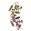
| Deposited unit | 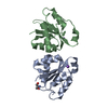
| ||||||||
|---|---|---|---|---|---|---|---|---|---|
| 1 |
| ||||||||
| Unit cell |
|
-Components
| #1: Protein | Mass: 14439.729 Da / Num. of mol.: 1 / Fragment: RECEIVER DOMAIN, RESIDUES 1-127 / Mutation: YES Source method: isolated from a genetically manipulated source Source: (gene. exp.) | ||||||||
|---|---|---|---|---|---|---|---|---|---|
| #2: Protein | Mass: 14411.716 Da / Num. of mol.: 1 / Fragment: RECEIVER DOMAIN, RESIDUES 1-127 / Mutation: YES Source method: isolated from a genetically manipulated source Source: (gene. exp.) | ||||||||
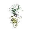
| #3: Chemical |   Mass: 22.990 Da / Num. of mol.: 2 / Source method: obtained synthetically / Formula: Na Mass: 22.990 Da / Num. of mol.: 2 / Source method: obtained synthetically / Formula: Na#4: Chemical | ChemComp-TRS / |   Mass: 122.143 Da / Num. of mol.: 1 / Source method: obtained synthetically / Formula: C4H12NO3 / Comment: pH buffer*YM Mass: 122.143 Da / Num. of mol.: 1 / Source method: obtained synthetically / Formula: C4H12NO3 / Comment: pH buffer*YM#5: Water | ChemComp-HOH / |  Mass: 18.015 Da / Num. of mol.: 303 / Source method: isolated from a natural source / Formula: H2O Mass: 18.015 Da / Num. of mol.: 303 / Source method: isolated from a natural source / Formula: H2OCompound details | ENGINEERED RESIDUE IN CHAIN A, ASP 53 TO ALA ENGINEERED RESIDUE IN CHAIN A, TYR 102 TO CYS ...ENGINEERED | Sequence details | MUTATIONS D53A,Y102C | |
-Experimental details
-Experiment
| Experiment | Method: X-RAY DIFFRACTION / Number of used crystals: 1 |
|---|
- Sample preparation
| Crystal | Density Matthews: 2.2 Å3/Da / Density % sol: 36.92 % |
|---|---|
| Crystal grow | pH: 8 Details: 3 MICROLITER OF PROTEIN SOLUTION AT 5.5 MG/ML AND 3 MICROLITER OF RESERVOIR SOLUTION (20% (W/V) PEG 4K, 0.4M SODIUM ACETATE, 0.1M TRISHCL (PH 8), 0.01M DTT |
-Data collection
| Diffraction | Mean temperature: 100 K |
|---|---|
| Diffraction source | Source: SYNCHROTRON / Site: ESRF  / Beamline: ID14-2 / Wavelength: 0.933 / Beamline: ID14-2 / Wavelength: 0.933 |
| Detector | Type: ADSC CCD / Detector: CCD |
| Radiation | Protocol: SINGLE WAVELENGTH / Monochromatic (M) / Laue (L): M / Scattering type: x-ray |
| Radiation wavelength | Wavelength: 0.933 Å / Relative weight: 1 |
| Reflection | Resolution: 1.45→58.7 Å / Num. obs: 41827 / % possible obs: 94.5 % / Observed criterion σ(I): 0 / Redundancy: 3.2 % / Rmerge(I) obs: 0.07 / Net I/σ(I): 15.2 |
| Reflection shell | Resolution: 1.45→1.53 Å / Redundancy: 3.2 % / Rmerge(I) obs: 0.29 / Mean I/σ(I) obs: 2.3 / % possible all: 70.3 |
- Processing
| Software |
| ||||||||||||||||||||||||||||||||||||||||||||||||||||||||||||||||||||||||||||||||||||||||||||||||||||||||||||||||||||||||||||||||||||||||||||||||||||||||||||||||||||||||||||||||||||||
|---|---|---|---|---|---|---|---|---|---|---|---|---|---|---|---|---|---|---|---|---|---|---|---|---|---|---|---|---|---|---|---|---|---|---|---|---|---|---|---|---|---|---|---|---|---|---|---|---|---|---|---|---|---|---|---|---|---|---|---|---|---|---|---|---|---|---|---|---|---|---|---|---|---|---|---|---|---|---|---|---|---|---|---|---|---|---|---|---|---|---|---|---|---|---|---|---|---|---|---|---|---|---|---|---|---|---|---|---|---|---|---|---|---|---|---|---|---|---|---|---|---|---|---|---|---|---|---|---|---|---|---|---|---|---|---|---|---|---|---|---|---|---|---|---|---|---|---|---|---|---|---|---|---|---|---|---|---|---|---|---|---|---|---|---|---|---|---|---|---|---|---|---|---|---|---|---|---|---|---|---|---|---|---|
| Refinement | Method to determine structure: MOLECULAR REPLACEMENT Starting model: PDB ENTRY 1B00 Resolution: 1.45→50 Å / Cor.coef. Fo:Fc: 0.961 / Cor.coef. Fo:Fc free: 0.957 / SU B: 1.224 / SU ML: 0.047 / TLS residual ADP flag: LIKELY RESIDUAL / Cross valid method: THROUGHOUT / ESU R: 0.074 / ESU R Free: 0.07 / Stereochemistry target values: MAXIMUM LIKELIHOOD / Details: HYDROGENS HAVE BEEN ADDED IN THE RIDING POSITIONS.
| ||||||||||||||||||||||||||||||||||||||||||||||||||||||||||||||||||||||||||||||||||||||||||||||||||||||||||||||||||||||||||||||||||||||||||||||||||||||||||||||||||||||||||||||||||||||
| Solvent computation | Ion probe radii: 0.8 Å / Shrinkage radii: 0.8 Å / VDW probe radii: 1.4 Å / Solvent model: BABINET MODEL WITH MASK | ||||||||||||||||||||||||||||||||||||||||||||||||||||||||||||||||||||||||||||||||||||||||||||||||||||||||||||||||||||||||||||||||||||||||||||||||||||||||||||||||||||||||||||||||||||||
| Displacement parameters | Biso mean: 12.69 Å2
| ||||||||||||||||||||||||||||||||||||||||||||||||||||||||||||||||||||||||||||||||||||||||||||||||||||||||||||||||||||||||||||||||||||||||||||||||||||||||||||||||||||||||||||||||||||||
| Refinement step | Cycle: LAST / Resolution: 1.45→50 Å
| ||||||||||||||||||||||||||||||||||||||||||||||||||||||||||||||||||||||||||||||||||||||||||||||||||||||||||||||||||||||||||||||||||||||||||||||||||||||||||||||||||||||||||||||||||||||
| Refine LS restraints |
|