 Movie
Movie Controller

[English] 日本語
 Yorodumi
- PDB-2jb2: The structure of L-amino acid oxidase from Rhodococcus opacus in ... -
+ Open data

- Basic information
| Entry | Database: PDB / ID: 2jb2 | ||||||
|---|---|---|---|---|---|---|---|
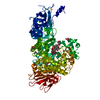
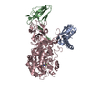
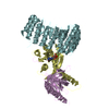
| Title | The structure of L-amino acid oxidase from Rhodococcus opacus in complex with L-phenylalanine. | ||||||
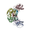
 Components Components | L-AMINO ACID OXIDASE | ||||||
 Keywords Keywords | OXIDOREDUCTASE / SUBSTRATE COMPLEX / L-AMINO ACID OXIDASE / NON-PLANAR FAD / DIMERISATION MODE / GR2-FAMILY / FLAVOENZYME / FAD CONTAINING / HYDRIDE TRANSFER MECHANISM | ||||||
| Function / homology |  Function and homology information Function and homology informationL-glutamate oxidase activity / L-lysine oxidase activity / L-phenylalaine oxidase activity / L-amino-acid oxidase / amino acid catabolic process / nucleotide binding / cytoplasm Similarity search - Function | ||||||
| Biological species |  RHODOCOCCUS OPACUS (bacteria) RHODOCOCCUS OPACUS (bacteria) | ||||||
| Method |  X-RAY DIFFRACTION / SYNCHROTRON / MOLECULAR REPLACEMENT / Resolution: 1.45 Å X-RAY DIFFRACTION / SYNCHROTRON / MOLECULAR REPLACEMENT / Resolution: 1.45 Å | ||||||
 Authors Authors | Faust, A. / Niefind, K. / Hummel, W. / Schomburg, D. | ||||||
 Citation Citation | Journal: J.Mol.Biol. / Year: 2007 Title: The Structure of a Bacterial L-Amino Acid Oxidase from Rhodococcus Opacus Gives New Evidence for the Hydride Mechanism for Dehydrogenation Authors: Faust, A. / Niefind, K. / Hummel, W. / Schomburg, D. #1: Journal: Acta Crystallogr.,Sect.F / Year: 2006 Title: Crystallization and Preliminary X-Ray Analysis of a Bacterial L-Amino-Acid Oxidase from Rhodococcus Opacus Authors: Faust, A. / Geueke, B. / Niefind, K. / Hummel, W. / Schomburg, D. | ||||||
| History |
|
- Structure visualization
| Structure viewer | Molecule: MolmilJmol/JSmol |
|---|
- Downloads & links
-Download
| PDBx/mmCIF format | 2jb2.cif.gz | 391.8 KB | Display | PDBx/mmCIF format |
|---|---|---|---|---|
| PDB format | pdb2jb2.ent.gz | 319.7 KB | Display | PDB format |
| PDBx/mmJSON format | 2jb2.json.gz | Tree view | PDBx/mmJSON format | |
| Others |  Other downloads Other downloads |
-Validation report
| Summary document | 2jb2_validation.pdf.gz | 930.8 KB | Display | wwPDB validaton report |
|---|---|---|---|---|
| Full document | 2jb2_full_validation.pdf.gz | 940.7 KB | Display | |
| Data in XML | 2jb2_validation.xml.gz | 41.9 KB | Display | |
| Data in CIF | 2jb2_validation.cif.gz | 62.3 KB | Display | |
| Arichive directory | https://data.pdbj.org/pub/pdb/validation_reports/jb/2jb2ftp://data.pdbj.org/pub/pdb/validation_reports/jb/2jb2 | HTTPS FTP |
-Related structure data
| Related structure data | 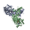 2jaeSC 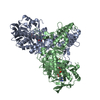 2jb1C  2jb3C S: Starting model for refinement C: citing same article ( |
|---|---|
| Similar structure data |


-Links
 PDBj
PDBj


- Assembly
| Deposited unit | 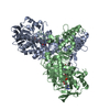
| ||||||||
|---|---|---|---|---|---|---|---|---|---|
| 1 |
| ||||||||
| Unit cell |
|
-Components
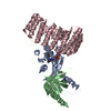
| #1: Protein | Mass: 53411.957 Da / Num. of mol.: 2 / Fragment: RESIDUES 46-534 / Source method: isolated from a natural source / Details: DSM 43250 / Source: (natural) RHODOCOCCUS OPACUS (bacteria) / References: UniProt: Q8VPD4, L-amino-acid oxidase#2: Chemical |   Type: L-peptide linking / Mass: 165.189 Da / Num. of mol.: 2 / Source method: obtained synthetically / Formula: C9H11NO2 Type: L-peptide linking / Mass: 165.189 Da / Num. of mol.: 2 / Source method: obtained synthetically / Formula: C9H11NO2#3: Chemical |   Mass: 785.550 Da / Num. of mol.: 2 / Source method: obtained synthetically / Formula: C27H33N9O15P2 / Comment: FAD*YM Mass: 785.550 Da / Num. of mol.: 2 / Source method: obtained synthetically / Formula: C27H33N9O15P2 / Comment: FAD*YM#4: Water | ChemComp-HOH / |  Mass: 18.015 Da / Num. of mol.: 711 / Source method: isolated from a natural source / Formula: H2O Mass: 18.015 Da / Num. of mol.: 711 / Source method: isolated from a natural source / Formula: H2O |
|---|
-Experimental details
-Experiment
| Experiment | Method: X-RAY DIFFRACTION / Number of used crystals: 1 |
|---|
- Sample preparation
| Crystal | Density Matthews: 2.4 Å3/Da / Density % sol: 40 % |
|---|---|
| Crystal grow | pH: 7.8 / Details: 100MM HEPES PH 7.8 10% 2-PROPANOLE 10% PEG 4000 |
-Data collection
| Diffraction | Mean temperature: 100 K |
|---|---|
| Diffraction source | Source: SYNCHROTRON / Site: BESSY  / Beamline: 14.2 / Wavelength: 0.9195 / Beamline: 14.2 / Wavelength: 0.9195 |
| Detector | Type: MARRESEARCH / Detector: IMAGE PLATE / Date: Jul 1, 2004 |
| Radiation | Protocol: SINGLE WAVELENGTH / Monochromatic (M) / Laue (L): M / Scattering type: x-ray |
| Radiation wavelength | Wavelength: 0.9195 Å / Relative weight: 1 |
| Reflection | Resolution: 1.45→30 Å / Num. obs: 157853 / % possible obs: 91.3 % / Redundancy: 3.2 % / Rmerge(I) obs: 0.1 / Net I/σ(I): 11.9 |
| Reflection shell | Resolution: 1.45→1.5 Å / Redundancy: 2.9 % / Rmerge(I) obs: 0.48 / Mean I/σ(I) obs: 2.1 / % possible all: 86.1 |
- Processing
| Software |
| ||||||||||||||||||||||||||||||||||||||||||||||||||||||||||||||||||||||||||||||||||||||||||||||||||||||||||||||||||||||||||||||||||||||||||||||||||||||||||||||||||||||||||||||||||||||
|---|---|---|---|---|---|---|---|---|---|---|---|---|---|---|---|---|---|---|---|---|---|---|---|---|---|---|---|---|---|---|---|---|---|---|---|---|---|---|---|---|---|---|---|---|---|---|---|---|---|---|---|---|---|---|---|---|---|---|---|---|---|---|---|---|---|---|---|---|---|---|---|---|---|---|---|---|---|---|---|---|---|---|---|---|---|---|---|---|---|---|---|---|---|---|---|---|---|---|---|---|---|---|---|---|---|---|---|---|---|---|---|---|---|---|---|---|---|---|---|---|---|---|---|---|---|---|---|---|---|---|---|---|---|---|---|---|---|---|---|---|---|---|---|---|---|---|---|---|---|---|---|---|---|---|---|---|---|---|---|---|---|---|---|---|---|---|---|---|---|---|---|---|---|---|---|---|---|---|---|---|---|---|---|
| Refinement | Method to determine structure: MOLECULAR REPLACEMENT Starting model: PDB ENTRY 2JAE Resolution: 1.45→14.99 Å / Cor.coef. Fo:Fc: 0.961 / Cor.coef. Fo:Fc free: 0.951 / SU B: 2.198 / SU ML: 0.04 / Cross valid method: THROUGHOUT / ESU R: 0.087 / ESU R Free: 0.071 Stereochemistry target values: MAXIMUM LIKELIHOODWITH PHASES Details: HYDROGENS HAVE BEEN ADDED IN THE RIDING POSITIONS. FAD IS NON-PLANAR
| ||||||||||||||||||||||||||||||||||||||||||||||||||||||||||||||||||||||||||||||||||||||||||||||||||||||||||||||||||||||||||||||||||||||||||||||||||||||||||||||||||||||||||||||||||||||
| Solvent computation | Ion probe radii: 0.8 Å / Shrinkage radii: 0.8 Å / VDW probe radii: 1.4 Å / Solvent model: MASK | ||||||||||||||||||||||||||||||||||||||||||||||||||||||||||||||||||||||||||||||||||||||||||||||||||||||||||||||||||||||||||||||||||||||||||||||||||||||||||||||||||||||||||||||||||||||
| Displacement parameters | Biso mean: 14.12 Å2
| ||||||||||||||||||||||||||||||||||||||||||||||||||||||||||||||||||||||||||||||||||||||||||||||||||||||||||||||||||||||||||||||||||||||||||||||||||||||||||||||||||||||||||||||||||||||
| Refinement step | Cycle: LAST / Resolution: 1.45→14.99 Å
| ||||||||||||||||||||||||||||||||||||||||||||||||||||||||||||||||||||||||||||||||||||||||||||||||||||||||||||||||||||||||||||||||||||||||||||||||||||||||||||||||||||||||||||||||||||||
| Refine LS restraints |
|