 Movie
Movie Controller

[English] 日本語
 Yorodumi
- PDB-2jab: A designed ankyrin repeat protein evolved to picomolar affinity t... -
+ Open data

- Basic information
| Entry | Database: PDB / ID: 2jab | ||||||
|---|---|---|---|---|---|---|---|
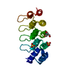
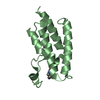
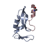
| Title | A designed ankyrin repeat protein evolved to picomolar affinity to Her2 | ||||||
 Components Components | H10-2-G3 | ||||||
 Keywords Keywords | DE NOVO PROTEIN / HER2 / DARPIN / ANKYRIN REPEAT PROTEIN / MEMBRANE PROTEIN / HUMAN EPIDERMAL GROWTH FACTOR RECEPTOR 2 | ||||||
| Function / homology | Ankyrin repeat-containing domain / Serine Threonine Protein Phosphatase 5, Tetratricopeptide repeat / Alpha Horseshoe / Mainly Alpha Function and homology information Function and homology information | ||||||
| Biological species | SYNTHETIC CONSTRUCT (others) | ||||||
| Method |  X-RAY DIFFRACTION / MOLECULAR REPLACEMENT / Resolution: 1.7 Å X-RAY DIFFRACTION / MOLECULAR REPLACEMENT / Resolution: 1.7 Å | ||||||
 Authors Authors | Zahnd, C. / Wyler, E. / Schwenk, J.M. / Steiner, D. / Lawrence, M.C. / McKern, N.M. / Pecorari, F. / Ward, C.W. / Joos, T.O. / Pluckthun, A. | ||||||
 Citation Citation | Journal: J.Mol.Biol. / Year: 2007 Title: A Designed Ankyrin Repeat Protein Evolved to Picomolar Affinity to Her2 Authors: Zahnd, C. / Wyler, E. / Schwenk, J.M. / Steiner, D. / Lawrence, M.C. / Mckern, N.M. / Pecorari, F. / Ward, C.W. / Joos, T.O. / Pluckthun, A. | ||||||
| History |
|
- Structure visualization
| Structure viewer | Molecule: MolmilJmol/JSmol |
|---|
- Downloads & links
-Download
| PDBx/mmCIF format | 2jab.cif.gz | 90 KB | Display | PDBx/mmCIF format |
|---|---|---|---|---|
| PDB format | pdb2jab.ent.gz | 70.4 KB | Display | PDB format |
| PDBx/mmJSON format | 2jab.json.gz | Tree view | PDBx/mmJSON format | |
| Others |  Other downloads Other downloads |
-Validation report
| Arichive directory | https://data.pdbj.org/pub/pdb/validation_reports/ja/2jabftp://data.pdbj.org/pub/pdb/validation_reports/ja/2jab | HTTPS FTP |
|---|
-Related structure data


-Links
 PDBj
PDBj
- Assembly
| Deposited unit | 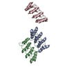
| ||||||||
|---|---|---|---|---|---|---|---|---|---|
| 1 | 
| ||||||||
| 2 | 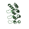
| ||||||||
| 3 | 
| ||||||||
| Unit cell |
| ||||||||
| Components on special symmetry positions |
|
-Components
| #1: Protein | Mass: 14618.437 Da / Num. of mol.: 3 Source method: isolated from a genetically manipulated source Source: (gene. exp.) SYNTHETIC CONSTRUCT (others) / Production host:  #2: Water | ChemComp-HOH / |  Mass: 18.015 Da / Num. of mol.: 488 / Source method: isolated from a natural source / Formula: H2O Mass: 18.015 Da / Num. of mol.: 488 / Source method: isolated from a natural source / Formula: H2O |
|---|
-Experimental details
-Experiment
| Experiment | Method: X-RAY DIFFRACTION / Number of used crystals: 1 |
|---|
- Sample preparation
| Crystal | Density Matthews: 2.88 Å3/Da / Density % sol: 57 % |
|---|---|
| Crystal grow | pH: 8.5 Details: PROTEIN: 6 MG/ML IN 20 MM SODIUM PHOSPHATE, 75 MM NACL PH 7.4. PRECIPITANT: 0.1 M TRISHCL (PH 8.5), 2.3 M (NH4)2SO4, 10% GLYCEROL (V/V). ROOM TEMPERATURE. |
-Data collection
| Diffraction | Mean temperature: 100 K |
|---|---|
| Diffraction source | Source: ROTATING ANODE / Type: RIGAKU RUH3R / Wavelength: 1.5418 |
| Detector | Type: RIGAKU IMAGE PLATE / Detector: IMAGE PLATE / Date: Dec 10, 2004 / Details: AXCO CAPILLARY |
| Radiation | Monochromator: NI FILTER / Protocol: SINGLE WAVELENGTH / Monochromatic (M) / Laue (L): M / Scattering type: x-ray |
| Radiation wavelength | Wavelength: 1.5418 Å / Relative weight: 1 |
| Reflection | Resolution: 1.7→73.3 Å / Num. obs: 58105 / % possible obs: 98.6 % / Redundancy: 3.8 % / Rmerge(I) obs: 0.12 / Net I/σ(I): 13.2 |
| Reflection shell | Resolution: 1.7→1.73 Å / Redundancy: 2.4 % / Rmerge(I) obs: 0.88 / Mean I/σ(I) obs: 1.3 / % possible all: 93.3 |
- Processing
| Software |
| ||||||||||||||||||||||||||||||||||||||||||||||||||||||||||||||||||||||||||||||||||||||||||||||||||||||||||||||||||||||||||||||||||||||||||||||||||||||||||||||||||||||||||||||||||||||
|---|---|---|---|---|---|---|---|---|---|---|---|---|---|---|---|---|---|---|---|---|---|---|---|---|---|---|---|---|---|---|---|---|---|---|---|---|---|---|---|---|---|---|---|---|---|---|---|---|---|---|---|---|---|---|---|---|---|---|---|---|---|---|---|---|---|---|---|---|---|---|---|---|---|---|---|---|---|---|---|---|---|---|---|---|---|---|---|---|---|---|---|---|---|---|---|---|---|---|---|---|---|---|---|---|---|---|---|---|---|---|---|---|---|---|---|---|---|---|---|---|---|---|---|---|---|---|---|---|---|---|---|---|---|---|---|---|---|---|---|---|---|---|---|---|---|---|---|---|---|---|---|---|---|---|---|---|---|---|---|---|---|---|---|---|---|---|---|---|---|---|---|---|---|---|---|---|---|---|---|---|---|---|---|
| Refinement | Method to determine structure: MOLECULAR REPLACEMENT Starting model: PDB ENTRIES 1MJ0 AND 2BKG Resolution: 1.7→15 Å / Cor.coef. Fo:Fc: 0.966 / Cor.coef. Fo:Fc free: 0.952 / SU B: 3.886 / SU ML: 0.065 / Cross valid method: THROUGHOUT / ESU R: 0.088 / ESU R Free: 0.092 / Stereochemistry target values: MAXIMUM LIKELIHOOD Details: HYDROGENS HAVE BEEN ADDED IN THE RIDING POSITIONS. NO NCS RESTRAINTS WERE APPLIED DURING THE FINAL STAGES OF REFINEMENT
| ||||||||||||||||||||||||||||||||||||||||||||||||||||||||||||||||||||||||||||||||||||||||||||||||||||||||||||||||||||||||||||||||||||||||||||||||||||||||||||||||||||||||||||||||||||||
| Solvent computation | Ion probe radii: 0.8 Å / Shrinkage radii: 0.8 Å / VDW probe radii: 1.2 Å / Solvent model: MASK | ||||||||||||||||||||||||||||||||||||||||||||||||||||||||||||||||||||||||||||||||||||||||||||||||||||||||||||||||||||||||||||||||||||||||||||||||||||||||||||||||||||||||||||||||||||||
| Displacement parameters | Biso mean: 35.07 Å2
| ||||||||||||||||||||||||||||||||||||||||||||||||||||||||||||||||||||||||||||||||||||||||||||||||||||||||||||||||||||||||||||||||||||||||||||||||||||||||||||||||||||||||||||||||||||||
| Refinement step | Cycle: LAST / Resolution: 1.7→15 Å
| ||||||||||||||||||||||||||||||||||||||||||||||||||||||||||||||||||||||||||||||||||||||||||||||||||||||||||||||||||||||||||||||||||||||||||||||||||||||||||||||||||||||||||||||||||||||
| Refine LS restraints |
|