 Movie
Movie Controller

[English] 日本語
 Yorodumi
- PDB-2j73: alpha-glucan rcognition by a family 41 carbohydrate-binding modul... -
+ Open data

- Basic information
| Entry | Database: PDB / ID: 2j73 | |||||||||
|---|---|---|---|---|---|---|---|---|---|---|
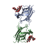
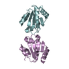
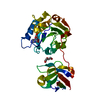
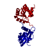
| Title | alpha-glucan rcognition by a family 41 carbohydrate-binding module from Thermotoga maritima pullulanase PulA | |||||||||
 Components Components | PULLULANASE | |||||||||
 Keywords Keywords | HYDROLASE / THERMOTOGA MARITIMA / ALPHA-GLUCAN BINDING / GLUCOSYL-MALTOTRIOSE / CARBOHYDRATE-BINDING MODULE / GLYCOSIDASE / BETA-SANDWICH FOLD | |||||||||
| Function / homology |  Function and homology information Function and homology informationpullulanase / pullulanase activity / carbohydrate binding / carbohydrate metabolic process Similarity search - Function | |||||||||
| Biological species |   THERMOTOGA MARITIMA (bacteria) THERMOTOGA MARITIMA (bacteria) | |||||||||
| Method |  X-RAY DIFFRACTION / MOLECULAR REPLACEMENT / Resolution: 1.4 Å X-RAY DIFFRACTION / MOLECULAR REPLACEMENT / Resolution: 1.4 Å | |||||||||
 Authors Authors | Lammerts van Bueren, A. / Boraston, A.B. | |||||||||
 Citation Citation | Journal: J.Mol.Biol. / Year: 2007 Title: The Structural Basis of Alpha-Glucan Recognition by a Family 41 Carbohydrate-Binding Module from Thermotoga Maritima Authors: Lammerts Van Bueren, A. / Boraston, A.B. | |||||||||
| History |
|
- Structure visualization
| Structure viewer | Molecule: MolmilJmol/JSmol |
|---|
- Downloads & links
-Download
| PDBx/mmCIF format | 2j73.cif.gz | 116.1 KB | Display | PDBx/mmCIF format |
|---|---|---|---|---|
| PDB format | pdb2j73.ent.gz | 90.5 KB | Display | PDB format |
| PDBx/mmJSON format | 2j73.json.gz | Tree view | PDBx/mmJSON format | |
| Others |  Other downloads Other downloads |
-Validation report
| Arichive directory | https://data.pdbj.org/pub/pdb/validation_reports/j7/2j73ftp://data.pdbj.org/pub/pdb/validation_reports/j7/2j73 | HTTPS FTP |
|---|
-Related structure data
| Related structure data | 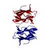 2j71SC  2j72C S: Starting model for refinement C: citing same article ( |
|---|---|
| Similar structure data |


-Links
 PDBj
PDBj

- Assembly
| Deposited unit | 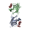
| |||||||||
|---|---|---|---|---|---|---|---|---|---|---|
| 1 |
| |||||||||
| Unit cell |
| |||||||||
| Components on special symmetry positions |
|
-Components
| #1: Protein | Mass: 12077.711 Da / Num. of mol.: 2 / Fragment: RESIDUES 20-120 Source method: isolated from a genetically manipulated source Source: (gene. exp.) THERMOTOGA MARITIMA (bacteria) / Plasmid: PET150 / Production host: #2: Polysaccharide | alpha-D-glucopyranose-(1-6)-alpha-D-glucopyranose-(1-4)-alpha-D-glucopyranose-(1-4)-alpha-D-glucopyranose | Source method: isolated from a genetically manipulated source #3: Polysaccharide | alpha-D-glucopyranose-(1-4)-alpha-D-glucopyranose-(1-4)-alpha-D-glucopyranose / alpha-maltotriose |   Source method: isolated from a genetically manipulated source Details: oligosaccharide / References: alpha-maltotriose #4: Water | ChemComp-HOH / |  Mass: 18.015 Da / Num. of mol.: 336 / Source method: isolated from a natural source / Formula: H2O Mass: 18.015 Da / Num. of mol.: 336 / Source method: isolated from a natural source / Formula: H2O |
|---|
-Experimental details
-Experiment
| Experiment | Method: X-RAY DIFFRACTION |
|---|
- Sample preparation
| Crystal | Density Matthews: 1.67 Å3/Da / Density % sol: 25.9 % |
|---|
-Data collection
| Diffraction | Mean temperature: 113 K |
|---|---|
| Diffraction source | Source: ROTATING ANODE / Wavelength: 1.5418 |
| Detector | Type: RIGAKU IMAGE PLATE / Detector: IMAGE PLATE |
| Radiation | Protocol: SINGLE WAVELENGTH / Monochromatic (M) / Laue (L): M / Scattering type: x-ray |
| Radiation wavelength | Wavelength: 1.5418 Å / Relative weight: 1 |
| Reflection | Resolution: 1.4→19.8 Å / Num. obs: 35936 / % possible obs: 91.2 % / Observed criterion σ(I): 2 / Redundancy: 3.65 % / Rmerge(I) obs: 0.04 / Net I/σ(I): 17.8 |
| Reflection shell | Resolution: 1.4→1.45 Å / Redundancy: 2.74 % / Rmerge(I) obs: 0.26 / Mean I/σ(I) obs: 2.8 / % possible all: 68.4 |
- Processing
| Software | Name: REFMAC / Version: 5.1.24 / Classification: refinement | ||||||||||||||||||||||||||||||||||||||||||||||||||||||||||||||||||||||||||||||||||||||||||||||||||||||||||||||||||||||||||||||||||||||||||||||||||||||||||||||||||||||||||||||||||||||
|---|---|---|---|---|---|---|---|---|---|---|---|---|---|---|---|---|---|---|---|---|---|---|---|---|---|---|---|---|---|---|---|---|---|---|---|---|---|---|---|---|---|---|---|---|---|---|---|---|---|---|---|---|---|---|---|---|---|---|---|---|---|---|---|---|---|---|---|---|---|---|---|---|---|---|---|---|---|---|---|---|---|---|---|---|---|---|---|---|---|---|---|---|---|---|---|---|---|---|---|---|---|---|---|---|---|---|---|---|---|---|---|---|---|---|---|---|---|---|---|---|---|---|---|---|---|---|---|---|---|---|---|---|---|---|---|---|---|---|---|---|---|---|---|---|---|---|---|---|---|---|---|---|---|---|---|---|---|---|---|---|---|---|---|---|---|---|---|---|---|---|---|---|---|---|---|---|---|---|---|---|---|---|---|
| Refinement | Method to determine structure: MOLECULAR REPLACEMENT Starting model: PDB ENTRY 2J71 Resolution: 1.4→50.64 Å / Cor.coef. Fo:Fc: 0.973 / Cor.coef. Fo:Fc free: 0.958 / SU B: 1.272 / SU ML: 0.049 / Cross valid method: THROUGHOUT / ESU R: 0.083 / ESU R Free: 0.076 / Stereochemistry target values: MAXIMUM LIKELIHOOD / Details: HYDROGENS HAVE BEEN ADDED IN THE RIDING POSITIONS.
| ||||||||||||||||||||||||||||||||||||||||||||||||||||||||||||||||||||||||||||||||||||||||||||||||||||||||||||||||||||||||||||||||||||||||||||||||||||||||||||||||||||||||||||||||||||||
| Solvent computation | Ion probe radii: 0.8 Å / Shrinkage radii: 0.8 Å / VDW probe radii: 1.4 Å / Solvent model: BABINET MODEL WITH MASK | ||||||||||||||||||||||||||||||||||||||||||||||||||||||||||||||||||||||||||||||||||||||||||||||||||||||||||||||||||||||||||||||||||||||||||||||||||||||||||||||||||||||||||||||||||||||
| Displacement parameters | Biso mean: 15.32 Å2
| ||||||||||||||||||||||||||||||||||||||||||||||||||||||||||||||||||||||||||||||||||||||||||||||||||||||||||||||||||||||||||||||||||||||||||||||||||||||||||||||||||||||||||||||||||||||
| Refinement step | Cycle: LAST / Resolution: 1.4→50.64 Å
| ||||||||||||||||||||||||||||||||||||||||||||||||||||||||||||||||||||||||||||||||||||||||||||||||||||||||||||||||||||||||||||||||||||||||||||||||||||||||||||||||||||||||||||||||||||||
| Refine LS restraints |
|