 Movie
Movie Controller


+ Open data

- Basic information
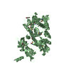
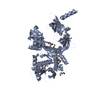
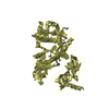
| Entry | Database: PDB / ID: 2ix0 | ||||||
|---|---|---|---|---|---|---|---|
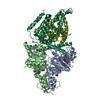
| Title | RNase II | ||||||
 Components Components | EXORIBONUCLEASE 2 | ||||||
 Keywords Keywords | HYDROLASE / S1 / RNA / CSD / RNB / NUCLEASE / RNASE II / RNA- BINDING / EXONUCLEASE | ||||||
| Function / homology |  Function and homology information Function and homology informationexoribonuclease II / exoribonuclease II activity / tRNA decay / mRNA catabolic process / 3'-5' exonuclease activity / RNA binding / cytosol Similarity search - Function | ||||||
| Biological species |  | ||||||
| Method |  X-RAY DIFFRACTION / SYNCHROTRON / MOLECULAR REPLACEMENT / Resolution: 2.44 Å X-RAY DIFFRACTION / SYNCHROTRON / MOLECULAR REPLACEMENT / Resolution: 2.44 Å | ||||||
 Authors Authors | Frazao, C. / Mcvey, C.E. / Amblar, M. / Barbas, A. / Vonrhein, C. / Arraiano, C.M. / Carrondo, M.A. | ||||||
 Citation Citation | Journal: Nature / Year: 2006 Title: Unravelling the Dynamics of RNA Degradation by Ribonuclease II and its RNA-Bound Complex Authors: Frazao, C. / Mcvey, C.E. / Amblar, M. / Barbas, A. / Vonrhein, C. / Arraiano, C.M. / Carrondo, M.A. #1: Journal: Acta Crystallogr.,Sect.F / Year: 2006 Title: Expression, Purification, Crystallization and Preliminary Diffraction Data Characterization of Escherichia Coli Ribonuclease II (Rnase II) Authors: Mcvey, C.E. / Amblar, M. / Barbas, A. / Cairrao, F. / Coelho, R. / Romao, C. / Arraiano, C.M. / Carrondo, M.A. / Frazao, C. #2: Journal: J.Mol.Biol. / Year: 2006 Title: Characterization of the Functional Domains of Escherichia Coli Rnase II. Authors: Amblar, M. / Barbas, A. / Fialho, A.M. / Arraiano, C.M. #3: Journal: Mol.Microbiol. / Year: 1993 Title: DNA Sequencing and Expression of the Gene Rnb Encoding Escherichia Coli Ribonuclease II Authors: Zilhao, R. / Camelo, L. / Arraiano, C.M. | ||||||
| History |
| ||||||
| Remark 700 | SHEET DETERMINATION METHOD: DSSP THE SHEETS PRESENTED AS "AA" IN EACH CHAIN ON SHEET RECORDS BELOW ... SHEET DETERMINATION METHOD: DSSP THE SHEETS PRESENTED AS "AA" IN EACH CHAIN ON SHEET RECORDS BELOW IS ACTUALLY AN 6-STRANDED BARREL THIS IS REPRESENTED BY A 7-STRANDED SHEET IN WHICH THE FIRST AND LAST STRANDS ARE IDENTICAL. |
- Structure visualization
| Structure viewer | Molecule: MolmilJmol/JSmol |
|---|
- Downloads & links
-Download
| PDBx/mmCIF format | 2ix0.cif.gz | 151 KB | Display | PDBx/mmCIF format |
|---|---|---|---|---|
| PDB format | pdb2ix0.ent.gz | 115.4 KB | Display | PDB format |
| PDBx/mmJSON format | 2ix0.json.gz | Tree view | PDBx/mmJSON format | |
| Others |  Other downloads Other downloads |
-Validation report
| Arichive directory | https://data.pdbj.org/pub/pdb/validation_reports/ix/2ix0ftp://data.pdbj.org/pub/pdb/validation_reports/ix/2ix0 | HTTPS FTP |
|---|
-Related structure data
| Related structure data | 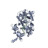 2ix1SC S: Starting model for refinement C: citing same article ( |
|---|---|
| Similar structure data |


-Links
 PDBj
PDBj


- Assembly
| Deposited unit | 
| ||||||||
|---|---|---|---|---|---|---|---|---|---|
| 1 |
| ||||||||
| Unit cell |
|
-Components
| #1: Protein | Mass: 74598.945 Da / Num. of mol.: 1 / Fragment: RESIDUES 6-644 Source method: isolated from a genetically manipulated source Source: (gene. exp.) |
|---|---|
| #2: Chemical | ChemComp-C5P /   Mass: 323.197 Da / Num. of mol.: 1 / Source method: obtained synthetically / Formula: C9H14N3O8P Mass: 323.197 Da / Num. of mol.: 1 / Source method: obtained synthetically / Formula: C9H14N3O8P |
| #3: Chemical | ChemComp-MG /   Mass: 24.305 Da / Num. of mol.: 1 / Source method: obtained synthetically / Formula: Mg Mass: 24.305 Da / Num. of mol.: 1 / Source method: obtained synthetically / Formula: Mg |
| #4: Chemical | ChemComp-CA /   Mass: 40.078 Da / Num. of mol.: 1 / Source method: obtained synthetically / Formula: Ca Mass: 40.078 Da / Num. of mol.: 1 / Source method: obtained synthetically / Formula: Ca |
| #5: Water | ChemComp-HOH /  Mass: 18.015 Da / Num. of mol.: 145 / Source method: isolated from a natural source / Formula: H2O Mass: 18.015 Da / Num. of mol.: 145 / Source method: isolated from a natural source / Formula: H2O |
| Sequence details | N-TERMINAL HIS-TAGGED PROTEIN, WHERE THE FIRST 5 RESIDUES, MFQDN, ARE REPLACED BY ...N-TERMINAL HIS-TAGGED PROTEIN, WHERE THE FIRST 5 RESIDUES, MFQDN, ARE REPLACED BY MGSSHHHHHH |
-Experimental details
-Experiment
| Experiment | Method: X-RAY DIFFRACTION / Number of used crystals: 1 |
|---|
- Sample preparation
| Crystal | Density Matthews: 3.13 Å3/Da / Density % sol: 60.73 % |
|---|---|
| Crystal grow | Temperature: 293 K / Method: vapor diffusion, sitting drop / pH: 8.5 Details: VAPOUR DIFFUSION IN SITTING DROPS WITH 1.5 MICRO-L OF PROTEIN SOLUTION, 8-12 MG/ML 3% GLYCEROL 1 MM MGCL2 100 MM KCL AND 100 MM TRIS-HCL PH 8.0, AND 1.5 MICRO-L OF WELL SOLUTION, 22 % PEG ...Details: VAPOUR DIFFUSION IN SITTING DROPS WITH 1.5 MICRO-L OF PROTEIN SOLUTION, 8-12 MG/ML 3% GLYCEROL 1 MM MGCL2 100 MM KCL AND 100 MM TRIS-HCL PH 8.0, AND 1.5 MICRO-L OF WELL SOLUTION, 22 % PEG 4K, 0.6M CA ACETATE AND 0.1M TRIS-HCL PH 8.5, AT 293 K |
-Data collection
| Diffraction | Mean temperature: 100 K |
|---|---|
| Diffraction source | Source: SYNCHROTRON / Site: ESRF  / Beamline: ID13 / Wavelength: 0.975 / Beamline: ID13 / Wavelength: 0.975 |
| Detector | Type: MARRESEARCH / Detector: CCD / Date: Mar 7, 2003 |
| Radiation | Protocol: SINGLE WAVELENGTH / Monochromatic (M) / Laue (L): M / Scattering type: x-ray |
| Radiation wavelength | Wavelength: 0.975 Å / Relative weight: 1 |
| Reflection | Resolution: 2.44→43.94 Å / Num. obs: 31569 / % possible obs: 98.9 % / Observed criterion σ(I): 0 / Redundancy: 7.3 % / Rmerge(I) obs: 0.08 / Net I/σ(I): 23.4 |
| Reflection shell | Resolution: 2.44→2.53 Å / Rmerge(I) obs: 0.42 / Mean I/σ(I) obs: 5.2 / % possible all: 98.2 |
- Processing
| Software |
| ||||||||||||||||||||||||||||||||||||||||||||||||||||||||||||||||||||||||||||||||||||||||||||||||||||||||||||||||||||||||||||||||||||||||||||||||||||||||||||||||||||||||||||||||||||||
|---|---|---|---|---|---|---|---|---|---|---|---|---|---|---|---|---|---|---|---|---|---|---|---|---|---|---|---|---|---|---|---|---|---|---|---|---|---|---|---|---|---|---|---|---|---|---|---|---|---|---|---|---|---|---|---|---|---|---|---|---|---|---|---|---|---|---|---|---|---|---|---|---|---|---|---|---|---|---|---|---|---|---|---|---|---|---|---|---|---|---|---|---|---|---|---|---|---|---|---|---|---|---|---|---|---|---|---|---|---|---|---|---|---|---|---|---|---|---|---|---|---|---|---|---|---|---|---|---|---|---|---|---|---|---|---|---|---|---|---|---|---|---|---|---|---|---|---|---|---|---|---|---|---|---|---|---|---|---|---|---|---|---|---|---|---|---|---|---|---|---|---|---|---|---|---|---|---|---|---|---|---|---|---|
| Refinement | Method to determine structure: MOLECULAR REPLACEMENT Starting model: PDB ENTRY 2IX1 Resolution: 2.44→62.87 Å / Cor.coef. Fo:Fc: 0.951 / Cor.coef. Fo:Fc free: 0.919 / SU B: 17.3 / SU ML: 0.196 / TLS residual ADP flag: LIKELY RESIDUAL / Cross valid method: THROUGHOUT / ESU R: 0.367 / ESU R Free: 0.249 / Stereochemistry target values: MAXIMUM LIKELIHOOD Details: HYDROGENS HAVE BEEN ADDED IN THE RIDING POSITIONS.DISOREDERED N-TERMINUS UNTIL RESIDUE 4 AND LOOP BETWEEN REIDUES 30-33 AND 67-70 WERE NOT MODELLED.
| ||||||||||||||||||||||||||||||||||||||||||||||||||||||||||||||||||||||||||||||||||||||||||||||||||||||||||||||||||||||||||||||||||||||||||||||||||||||||||||||||||||||||||||||||||||||
| Solvent computation | Ion probe radii: 0.8 Å / Shrinkage radii: 0.8 Å / VDW probe radii: 1.2 Å / Solvent model: BABINET MODEL WITH MASK | ||||||||||||||||||||||||||||||||||||||||||||||||||||||||||||||||||||||||||||||||||||||||||||||||||||||||||||||||||||||||||||||||||||||||||||||||||||||||||||||||||||||||||||||||||||||
| Displacement parameters | Biso mean: 39.85 Å2
| ||||||||||||||||||||||||||||||||||||||||||||||||||||||||||||||||||||||||||||||||||||||||||||||||||||||||||||||||||||||||||||||||||||||||||||||||||||||||||||||||||||||||||||||||||||||
| Refinement step | Cycle: LAST / Resolution: 2.44→62.87 Å
| ||||||||||||||||||||||||||||||||||||||||||||||||||||||||||||||||||||||||||||||||||||||||||||||||||||||||||||||||||||||||||||||||||||||||||||||||||||||||||||||||||||||||||||||||||||||
| Refine LS restraints |
|