[English] 日本語
Yorodumi
- PDB-2iuu: P. aeruginosa FtsK motor domain, hexamer -

+
Open data


ID or keywords:

Loading...

-
Basic information

Entry
Database: PDB / ID: 2iuu
TitleP. aeruginosa FtsK motor domain, hexamer
ComponentsDNA TRANSLOCASE FTSK
KeywordsMEMBRANE PROTEIN / DNA TRANSLOCATION / NUCLEOTIDE-BINDING / ATP-BINDING / DNA-BINDING / CELL DIVISION / TRANSMEMBRANE / CHROMOSOME PARTITION / INNER MEMBRANE / HEXAMERIC RING / KOPS / MEMBRANE / DIVISOME / CELL CYCLE / AAA ATPASE
Function / homology
Function and homology information


cellular response to antibiotic / chromosome segregation / cell division / DNA binding / ATP binding / plasma membrane
Similarity search - Function
Threonyl-tRNA Synthetase; Chain A, domain 2 - #40 / FtsK gamma domain / DNA translocase FtsK, 4TM region / FtsK alpha domain / Ftsk gamma domain / 4TM region of DNA translocase FtsK/SpoIIIE / FtsK alpha domain / Ftsk_gamma / Threonyl-tRNA Synthetase; Chain A, domain 2 / FtsK domain ...Threonyl-tRNA Synthetase; Chain A, domain 2 - #40 / FtsK gamma domain / DNA translocase FtsK, 4TM region / FtsK alpha domain / Ftsk gamma domain / 4TM region of DNA translocase FtsK/SpoIIIE / FtsK alpha domain / Ftsk_gamma / Threonyl-tRNA Synthetase; Chain A, domain 2 / FtsK domain / FtsK/SpoIIIE family / FtsK domain profile. / Winged helix DNA-binding domain superfamily / P-loop containing nucleotide triphosphate hydrolases / Winged helix-like DNA-binding domain superfamily / P-loop containing nucleoside triphosphate hydrolase / Rossmann fold / 2-Layer Sandwich / 3-Layer(aba) Sandwich / Alpha Beta
Similarity search - Domain/homology
ADENOSINE-5'-DIPHOSPHATE / DNA translocase FtsK
Similarity search - Component
Biological speciesPSEUDOMONAS AERUGINOSA (bacteria)
MethodX-RAY DIFFRACTION / SYNCHROTRON / MOLECULAR REPLACEMENT / Resolution: 2.9 Å
AuthorsMassey, T.H. / Mercogliano, C.P. / Yates, J. / Sherratt, D.J. / Lowe, J.
CitationJournal: Mol.Cell / Year: 2006
Title: Double-Stranded DNA Translocation: Structure and Mechanism of Hexameric Ftsk
Authors: Massey, T.H. / Mercogliano, C.P. / Yates, J. / Sherratt, D.J. / Lowe, J.
History
DepositionJun 7, 2006Deposition site: PDBE / Processing site: PDBE
Revision 1.0Aug 29, 2006Provider: repository / Type: Initial release
Revision 1.1May 8, 2011Group: Version format compliance
Revision 1.2Jul 13, 2011Group: Version format compliance

-
Structure visualization

Structure viewerMolecule:
MolmilJmol/JSmol

Downloads & links

-
Assembly

Deposited unit
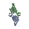
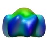
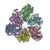
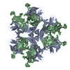
A: DNA TRANSLOCASE FTSK
B: DNA TRANSLOCASE FTSK
C: DNA TRANSLOCASE FTSK
D: DNA TRANSLOCASE FTSK
E: DNA TRANSLOCASE FTSK
F: DNA TRANSLOCASE FTSK
hetero molecules


Theoretical massNumber of molelcules
Total (without water)326,57212
Polymers324,0096
Non-polymers2,5636
Water0
1


  • Idetical with deposited unit
  • defined by author&software
TypeNameSymmetry operationNumber
identity operation1_555x,y,z1
MethodPQS
Unit cell
Length a, b, c (Å)140.012, 221.757, 134.075
Angle α, β, γ (deg.)90.00, 90.00, 90.00
Int Tables number18
Space group name H-MP21212

-
Components

#1: Protein
DNA TRANSLOCASE FTSK / FTSK MOTOR DOMAIN


Mass: 54001.441 Da / Num. of mol.: 6 / Fragment: MOTOR DOMAIN, RESIDUES 247-728
Source method: isolated from a genetically manipulated source
Source: (gene. exp.) PSEUDOMONAS AERUGINOSA (bacteria) / Production host: ESCHERICHIA COLI (E. coli) / References: UniProt: Q9I0M3
#2: Chemical
ChemComp-ADP / ADENOSINE-5'-DIPHOSPHATE / Adenosine diphosphate


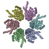
Mass: 427.201 Da / Num. of mol.: 6 / Source method: obtained synthetically / Formula: C10H15N5O10P2 / Comment: ADP, energy-carrying molecule*YM
Compound detailsDNA MOTOR PROTEIN, WHICH IS BOTH REQUIRED TO MOVE DNA OUT OF THE REGION OF THE SEPTUM DURING CELL ...DNA MOTOR PROTEIN, WHICH IS BOTH REQUIRED TO MOVE DNA OUT OF THE REGION OF THE SEPTUM DURING CELL DIVISION AND FOR THE SEPTUM FORMATION. TRACKS DNA IN AN ATP-DEPENDENT MANNER BY GENERATING POSITIVE SUPERCOILS IN FRONT OF IT AND NEGATIVE SUPERCOILS BEHIND IT (BY SIMILARITY).

-
Experimental details

-
Experiment

ExperimentMethod: X-RAY DIFFRACTION

-
Sample preparation

CrystalDensity Matthews: 3.47 Å3/Da / Density % sol: 65 %

-
Data collection

DiffractionMean temperature: 100 K
Diffraction sourceSource: SYNCHROTRON / Site: ESRF / Beamline: ID14-4 / Wavelength: 0.934
DetectorType: ADSC CCD / Detector: CCD
RadiationProtocol: SINGLE WAVELENGTH / Monochromatic (M) / Laue (L): M / Scattering type: x-ray
Radiation wavelengthWavelength: 0.934 Å / Relative weight: 1
ReflectionResolution: 2.9→100 Å / Num. obs: 90848 / % possible obs: 99.8 % / Observed criterion σ(I): 0 / Redundancy: 3.7 % / Biso Wilson estimate: 76 Å2 / Rmerge(I) obs: 0.08 / Net I/σ(I): 13.1
Reflection shellResolution: 2.9→3.06 Å / Redundancy: 3.7 % / Rmerge(I) obs: 0.38 / Mean I/σ(I) obs: 3.3 / % possible all: 99.8

-
Processing

Software
NameVersionClassification
CNS1.1refinement
MOSFLMdata reduction
SCALAdata scaling
PHASERphasing
RefinementMethod to determine structure: MOLECULAR REPLACEMENT / Resolution: 2.9→100 Å / Data cutoff high absF: 10000 / Cross valid method: THROUGHOUT / σ(F): 0 / Stereochemistry target values: MLF
RfactorNum. reflection% reflectionSelection details
Rfree0.2594 4650 5 %RANDOM
Rwork0.2334 ---
obs0.2334 92748 99.6 %-
Solvent computationBsol: 37.1892 Å2 / ksol: 0.33365 e/Å3
Displacement parametersBiso mean: 67 Å2
Baniso -1Baniso -2Baniso -3
1-7.078 Å20 Å20 Å2
2--9.913 Å20 Å2
3----16.991 Å2
Refinement stepCycle: LAST / Resolution: 2.9→100 Å
ProteinNucleic acidLigandSolventTotal
Num. atoms18726 0 162 0 18888
Refine LS restraints
Refine-IDTypeDev ideal
X-RAY DIFFRACTIONc_bond_d0.009
X-RAY DIFFRACTIONc_bond_d_na
X-RAY DIFFRACTIONc_bond_d_prot
X-RAY DIFFRACTIONc_angle_d
X-RAY DIFFRACTIONc_angle_d_na
X-RAY DIFFRACTIONc_angle_d_prot
X-RAY DIFFRACTIONc_angle_deg1.521
X-RAY DIFFRACTIONc_angle_deg_na
X-RAY DIFFRACTIONc_angle_deg_prot
X-RAY DIFFRACTIONc_dihedral_angle_d
X-RAY DIFFRACTIONc_dihedral_angle_d_na
X-RAY DIFFRACTIONc_dihedral_angle_d_prot
X-RAY DIFFRACTIONc_improper_angle_d
X-RAY DIFFRACTIONc_improper_angle_d_na
X-RAY DIFFRACTIONc_improper_angle_d_prot
X-RAY DIFFRACTIONc_mcbond_it
X-RAY DIFFRACTIONc_mcangle_it
X-RAY DIFFRACTIONc_scbond_it
X-RAY DIFFRACTIONc_scangle_it
LS refinement shellResolution: 2.9→2.92 Å / Rfactor Rfree: 0.3981 / Rfactor Rwork: 0.3721 / Total num. of bins used: 50
Xplor file
Refine-IDSerial noParam file
X-RAY DIFFRACTION1PROTEIN_REP.PARAM
X-RAY DIFFRACTION2ION.PARAM
X-RAY DIFFRACTION3WATER_REP.PARAM
X-RAY DIFFRACTION4ADP.PAR

+
About Yorodumi

-
News

-
Feb 9, 2022. New format data for meta-information of EMDB entries

New format data for meta-information of EMDB entries

  • Version 3 of the EMDB header file is now the official format.
  • The previous official version 1.9 will be removed from the archive.

Related info.:EMDB header

External links:wwPDB to switch to version 3 of the EMDB data model

-
Aug 12, 2020. Covid-19 info

Covid-19 info

URL: https://pdbj.org/emnavi/covid19.php

New page: Covid-19 featured information page in EM Navigator.

Related info.:Covid-19 info / Mar 5, 2020. Novel coronavirus structure data

+
Mar 5, 2020. Novel coronavirus structure data

Novel coronavirus structure data

Related info.:Yorodumi Speices / Aug 12, 2020. Covid-19 info

External links:COVID-19 featured content - PDBj / Molecule of the Month (242):Coronavirus Proteases

+
Jan 31, 2019. EMDB accession codes are about to change! (news from PDBe EMDB page)

EMDB accession codes are about to change! (news from PDBe EMDB page)

  • The allocation of 4 digits for EMDB accession codes will soon come to an end. Whilst these codes will remain in use, new EMDB accession codes will include an additional digit and will expand incrementally as the available range of codes is exhausted. The current 4-digit format prefixed with “EMD-” (i.e. EMD-XXXX) will advance to a 5-digit format (i.e. EMD-XXXXX), and so on. It is currently estimated that the 4-digit codes will be depleted around Spring 2019, at which point the 5-digit format will come into force.
  • The EM Navigator/Yorodumi systems omit the EMD- prefix.

Related info.:Q: What is EMD? / ID/Accession-code notation in Yorodumi/EM Navigator

External links:EMDB Accession Codes are Changing Soon! / Contact to PDBj

+
Jul 12, 2017. Major update of PDB

Major update of PDB

  • wwPDB released updated PDB data conforming to the new PDBx/mmCIF dictionary.
  • This is a major update changing the version number from 4 to 5, and with Remediation, in which all the entries are updated.
  • In this update, many items about electron microscopy experimental information are reorganized (e.g. em_software).
  • Now, EM Navigator and Yorodumi are based on the updated data.

External links:wwPDB Remediation / Enriched Model Files Conforming to OneDep Data Standards Now Available in the PDB FTP Archive

-
Yorodumi

Thousand views of thousand structures

  • Yorodumi is a browser for structure data from EMDB, PDB, SASBDB, etc.
  • This page is also the successor to EM Navigator detail page, and also detail information page/front-end page for Omokage search.
  • The word "yorodu" (or yorozu) is an old Japanese word meaning "ten thousand". "mi" (miru) is to see.

Related info.:EMDB / PDB / SASBDB / Comparison of 3 databanks / Yorodumi Search / Aug 31, 2016. New EM Navigator & Yorodumi / Yorodumi Papers / Jmol/JSmol / Function and homology information / Changes in new EM Navigator and Yorodumi

Read more