 Movie
Movie Controller

[English] 日本語
 Yorodumi
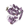
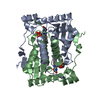
- PDB-2isl: BluB bound to reduced flavin (FMNH2) and molecular oxygen. (clear... -
+ Open data

- Basic information
| Entry | Database: PDB / ID: 2isl | ||||||
|---|---|---|---|---|---|---|---|
| Title | BluB bound to reduced flavin (FMNH2) and molecular oxygen. (clear crystal form) | ||||||
 Components Components | BluB | ||||||
 Keywords Keywords | FLAVOPROTEIN / flavin / oxidoreductase / monooxygenase / flavin destructase / molecular oxygen | ||||||
| Function / homology |  Function and homology information Function and homology informationaerobic 5,6-dimethylbenzimidazole synthase / 5,6-dimethylbenzimidazole synthase activity / cobalamin biosynthetic process / oxidoreductase activity, acting on paired donors, with incorporation or reduction of molecular oxygen / nucleotide binding Similarity search - Function | ||||||
| Biological species |  Sinorhizobium meliloti (bacteria) Sinorhizobium meliloti (bacteria) | ||||||
| Method |  X-RAY DIFFRACTION / SYNCHROTRON / MOLECULAR REPLACEMENT / Resolution: 2.9 Å X-RAY DIFFRACTION / SYNCHROTRON / MOLECULAR REPLACEMENT / Resolution: 2.9 Å | ||||||
 Authors Authors | Larsen, N.A. / Taga, M.E. / Howard-Jones, A.R. / Walsh, C.T. / Walker, G.C. | ||||||
 Citation Citation | Journal: Nature / Year: 2007 Title: BluB cannibalizes flavin to form the lower ligand of vitamin B12. Authors: Taga, M.E. / Larsen, N.A. / Howard-Jones, A.R. / Walsh, C.T. / Walker, G.C. #1: Journal: Proc.Natl.Acad.Sci.USA / Year: 2006 Title: Sinorhizobium meliloti bluB is necessary for production of 5,6-dimethylbenzimidazole, the lower ligand of B12. | ||||||
| History |
|
- Structure visualization
| Structure viewer | Molecule: MolmilJmol/JSmol |
|---|
- Downloads & links
-Download
| PDBx/mmCIF format | 2isl.cif.gz | 351.5 KB | Display | PDBx/mmCIF format |
|---|---|---|---|---|
| PDB format | pdb2isl.ent.gz | 289.5 KB | Display | PDB format |
| PDBx/mmJSON format | 2isl.json.gz | Tree view | PDBx/mmJSON format | |
| Others |  Other downloads Other downloads |
-Validation report
| Arichive directory | https://data.pdbj.org/pub/pdb/validation_reports/is/2islftp://data.pdbj.org/pub/pdb/validation_reports/is/2isl | HTTPS FTP |
|---|
-Related structure data


-Links
 PDBj
PDBj


- Assembly
| Deposited unit | 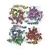
| ||||||||
|---|---|---|---|---|---|---|---|---|---|
| 1 | 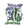
| ||||||||
| 2 | 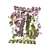
| ||||||||
| 3 | 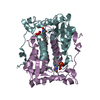
| ||||||||
| 4 | 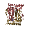
| ||||||||
| Unit cell |
| ||||||||
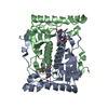
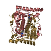
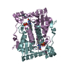
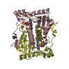
| Details | The biological assembly consists of a homodimer. There are 4 dimers in the asymmetric unit. Dimer1 = chain A/B, Dimer2 = chain C/D, Dimer3 = chain E/F, Dimer4 = chain G/H |
-Components
| #1: Protein | Mass: 25820.369 Da / Num. of mol.: 8 Source method: isolated from a genetically manipulated source Source: (gene. exp.) Sinorhizobium meliloti (bacteria) / Gene: bluB / Plasmid: pET-28b / Species (production host): Escherichia coli / Production host: #2: Chemical | ChemComp-FNR / 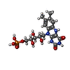  Mass: 458.360 Da / Num. of mol.: 8 / Source method: obtained synthetically / Formula: C17H23N4O9P Mass: 458.360 Da / Num. of mol.: 8 / Source method: obtained synthetically / Formula: C17H23N4O9P#3: Chemical | ChemComp-OXY /   Mass: 31.999 Da / Num. of mol.: 8 / Source method: obtained synthetically / Formula: O2 Mass: 31.999 Da / Num. of mol.: 8 / Source method: obtained synthetically / Formula: O2#4: Water | ChemComp-HOH / |  Mass: 18.015 Da / Num. of mol.: 63 / Source method: isolated from a natural source / Formula: H2O Mass: 18.015 Da / Num. of mol.: 63 / Source method: isolated from a natural source / Formula: H2O |
|---|
-Experimental details
-Experiment
| Experiment | Method: X-RAY DIFFRACTION / Number of used crystals: 1 |
|---|
- Sample preparation
| Crystal | Density Matthews: 2.52 Å3/Da / Density % sol: 51.14 % |
|---|---|
| Crystal grow | Temperature: 298 K / Method: vapor diffusion, hanging drop / pH: 5.6 Details: 0.8 Ammonium Sulfate, 100 mM citrate soaked in saturated dithionite, and then backsoaked in oxygenated mother liquor prior to freezing, pH 5.6, VAPOR DIFFUSION, HANGING DROP, temperature 298K |
-Data collection
| Diffraction | Mean temperature: 120 K |
|---|---|
| Diffraction source | Source: SYNCHROTRON / Site: ALS  / Beamline: 8.2.2 / Wavelength: 0.9919 Å / Beamline: 8.2.2 / Wavelength: 0.9919 Å |
| Detector | Type: ADSC QUANTUM 315 / Detector: CCD / Date: Jun 2, 2006 |
| Radiation | Protocol: SINGLE WAVELENGTH / Monochromatic (M) / Laue (L): M / Scattering type: x-ray |
| Radiation wavelength | Wavelength: 0.9919 Å / Relative weight: 1 |
| Reflection | Resolution: 2.9→50 Å / Num. all: 44966 / Num. obs: 44966 / % possible obs: 100 % / Observed criterion σ(F): 3.3 / Observed criterion σ(I): 3.3 / Redundancy: 3.8 % / Rmerge(I) obs: 0.079 / Net I/σ(I): 15.1 |
| Reflection shell | Resolution: 2.9→3 Å / Redundancy: 3.7 % / Rmerge(I) obs: 0.315 / Mean I/σ(I) obs: 3.3 / % possible all: 100 |
- Processing
| Software |
| ||||||||||||||||||||||||||||||||||||||||||||||||||||||||||||||||||||||||||||||||
|---|---|---|---|---|---|---|---|---|---|---|---|---|---|---|---|---|---|---|---|---|---|---|---|---|---|---|---|---|---|---|---|---|---|---|---|---|---|---|---|---|---|---|---|---|---|---|---|---|---|---|---|---|---|---|---|---|---|---|---|---|---|---|---|---|---|---|---|---|---|---|---|---|---|---|---|---|---|---|---|---|---|
| Refinement | Method to determine structure: MOLECULAR REPLACEMENT / Resolution: 2.9→20 Å / Isotropic thermal model: isotropic / Cross valid method: THROUGHOUT / σ(F): 0 / σ(I): 0 / Stereochemistry target values: Engh & Huber
| ||||||||||||||||||||||||||||||||||||||||||||||||||||||||||||||||||||||||||||||||
| Displacement parameters | Biso mean: 36 Å2
| ||||||||||||||||||||||||||||||||||||||||||||||||||||||||||||||||||||||||||||||||
| Refinement step | Cycle: LAST / Resolution: 2.9→20 Å
| ||||||||||||||||||||||||||||||||||||||||||||||||||||||||||||||||||||||||||||||||
| Refine LS restraints |
| ||||||||||||||||||||||||||||||||||||||||||||||||||||||||||||||||||||||||||||||||
| Xplor file |
|
