 Movie
Movie Controller

[English] 日本語
 Yorodumi
- PDB-2ihx: Solution Structure of the Rous Sarcoma Virus Nucleocapsid Protein... -
+ Open data

- Basic information
| Entry | Database: PDB / ID: 2ihx | ||||||
|---|---|---|---|---|---|---|---|
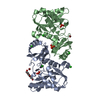
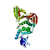
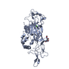
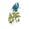
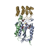
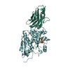
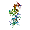
| Title | Solution Structure of the Rous Sarcoma Virus Nucleocapsid Protein:uPsi RNA Packaging Signal Complex | ||||||
 Components Components |
| ||||||
 Keywords Keywords | VIRAL PROTEIN/RNA / Protein-RNA complex / VIRAL PROTEIN-RNA COMPLEX | ||||||
| Function / homology |  Function and homology information Function and homology informationhost cell nucleoplasm / viral procapsid maturation / host cell nucleolus / Hydrolases; Acting on peptide bonds (peptidases); Aspartic endopeptidases / viral capsid / structural constituent of virion / aspartic-type endopeptidase activity / nucleic acid binding / viral translational frameshifting / host cell plasma membrane ...host cell nucleoplasm / viral procapsid maturation / host cell nucleolus / Hydrolases; Acting on peptide bonds (peptidases); Aspartic endopeptidases / viral capsid / structural constituent of virion / aspartic-type endopeptidase activity / nucleic acid binding / viral translational frameshifting / host cell plasma membrane / proteolysis / zinc ion binding Similarity search - Function | ||||||
| Biological species |  Rous sarcoma virus Rous sarcoma virus | ||||||
| Method | SOLUTION NMR / 1H-1H distance restraints, Hydrogen-bond restraints, Torsion angle restraints, Inter-phosphate restraints | ||||||
 Authors Authors | Zhou, J. / Summers, M. | ||||||
 Citation Citation | Journal: J.Mol.Biol. / Year: 2007 Title: Solution Structure of the Rous Sarcoma Virus Nucleocapsid Protein: muPsi RNA Packaging Signal Complex. Authors: Zhou, J. / Bean, R.L. / Vogt, V.M. / Summers, M. | ||||||
| History |
|
- Structure visualization
| Structure viewer | Molecule: MolmilJmol/JSmol |
|---|
- Downloads & links
-Download
| PDBx/mmCIF format | 2ihx.cif.gz | 1.4 MB | Display | PDBx/mmCIF format |
|---|---|---|---|---|
| PDB format | pdb2ihx.ent.gz | 1.2 MB | Display | PDB format |
| PDBx/mmJSON format | 2ihx.json.gz | Tree view | PDBx/mmJSON format | |
| Others |  Other downloads Other downloads |
-Validation report
| Arichive directory | https://data.pdbj.org/pub/pdb/validation_reports/ih/2ihxftp://data.pdbj.org/pub/pdb/validation_reports/ih/2ihx | HTTPS FTP |
|---|
-Related structure data
| Similar structure data |
|---|


-Links
 PDBj
PDBj


- Assembly
| Deposited unit | 
| |||||||||
|---|---|---|---|---|---|---|---|---|---|---|
| 1 |
| |||||||||
| NMR ensembles |
|
-Components
| #1: RNA chain | Mass: 24267.391 Da / Num. of mol.: 1 Fragment: minimal RNA packaging signal in the 5'- untranslated region (UTR) of Rous sarcoma virus (RSV) Source method: obtained synthetically Details: RNA was prepared by in vitro T7 RNA transcription. The sequence occurs naturally in Rous sarcoma virus (RSV) |
|---|---|
| #2: Protein | Mass: 6706.681 Da / Num. of mol.: 1 / Fragment: Nucleocapsid domain (residues 503-563) Source method: isolated from a genetically manipulated source Source: (gene. exp.) Rous sarcoma virus / Genus: Alpharetrovirus / Strain: Prague C (Pr-C) / Gene: GAG / Plasmid: pGEX-6P-1 / Production host:  |
| #3: Chemical |   Mass: 65.409 Da / Num. of mol.: 2 / Source method: obtained synthetically / Formula: Zn Mass: 65.409 Da / Num. of mol.: 2 / Source method: obtained synthetically / Formula: Zn |
-Experimental details
-Experiment
| Experiment | Method: SOLUTION NMR | ||||||||||||||||||||||||||||
|---|---|---|---|---|---|---|---|---|---|---|---|---|---|---|---|---|---|---|---|---|---|---|---|---|---|---|---|---|---|
| NMR experiment |
|
- Sample preparation
| Details |
| |||||||||||||||||||||
|---|---|---|---|---|---|---|---|---|---|---|---|---|---|---|---|---|---|---|---|---|---|---|
| Sample conditions | Ionic strength: 5 mM NaCl, 0.1 mM ZnCl2 / pH: 7 / Pressure: ambient / Temperature: 308 K |
-NMR measurement
| Radiation | Protocol: SINGLE WAVELENGTH / Monochromatic (M) / Laue (L): M | |||||||||||||||
|---|---|---|---|---|---|---|---|---|---|---|---|---|---|---|---|---|
| Radiation wavelength | Relative weight: 1 | |||||||||||||||
| NMR spectrometer |
|
- Processing
| NMR software |
| ||||||||||||||||||||
|---|---|---|---|---|---|---|---|---|---|---|---|---|---|---|---|---|---|---|---|---|---|
| Refinement | Method: 1H-1H distance restraints, Hydrogen-bond restraints, Torsion angle restraints, Inter-phosphate restraints Software ordinal: 1 Details: The structures are based on a total of 1697 restraints. 680 are NOE-derived distance restraints, 608 are Hydrogen-bond restraints, 239 are torsion angle restraints and 170 are inter-phosphate restraints. | ||||||||||||||||||||
| NMR representative | Selection criteria: fewest violations | ||||||||||||||||||||
| NMR ensemble | Conformer selection criteria: structures with the least restraint violations Conformers calculated total number: 800 / Conformers submitted total number: 20 |
 NMRPipe