 Movie
Movie Controller

[English] 日本語
 Yorodumi
- PDB-2ihf: Crystal structure of deletion mutant delta 228-252 R190A of the s... -
+ Open data

- Basic information
| Entry | Database: PDB / ID: 2ihf | ||||||
|---|---|---|---|---|---|---|---|
| Title | Crystal structure of deletion mutant delta 228-252 R190A of the single-stranded DNA binding protein from Thermus aquaticus | ||||||
 Components Components | Single-stranded DNA-binding protein | ||||||
 Keywords Keywords | DNA BINDING PROTEIN / Single-stranded DNA binding protein (SSB) / Thermophile organism / Protein-DNA interaction / Protein-protein interaction | ||||||
| Function / homology |  Function and homology information Function and homology informationnucleoid / single-stranded DNA binding / DNA recombination / DNA replication / DNA repair Similarity search - Function | ||||||
| Biological species |   Thermus aquaticus (bacteria) Thermus aquaticus (bacteria) | ||||||
| Method |  X-RAY DIFFRACTION / SYNCHROTRON / MOLECULAR REPLACEMENT / Resolution: 1.9 Å X-RAY DIFFRACTION / SYNCHROTRON / MOLECULAR REPLACEMENT / Resolution: 1.9 Å | ||||||
 Authors Authors | Fedorov, R. / Witte, G. / Urbanke, C. / Manstein, D.J. / Curth, U. | ||||||
 Citation Citation | Journal: Nucleic Acids Res. / Year: 2006 Title: 3D structure of Thermus aquaticus single-stranded DNA-binding protein gives insight into the functioning of SSB proteins. Authors: Fedorov, R. / Witte, G. / Urbanke, C. / Manstein, D.J. / Curth, U. | ||||||
| History |
|
- Structure visualization
| Structure viewer | Molecule: MolmilJmol/JSmol |
|---|
- Downloads & links
-Download
| PDBx/mmCIF format | 2ihf.cif.gz | 59.5 KB | Display | PDBx/mmCIF format |
|---|---|---|---|---|
| PDB format | pdb2ihf.ent.gz | 42.7 KB | Display | PDB format |
| PDBx/mmJSON format | 2ihf.json.gz | Tree view | PDBx/mmJSON format | |
| Others |  Other downloads Other downloads |
-Validation report
| Arichive directory | https://data.pdbj.org/pub/pdb/validation_reports/ih/2ihfftp://data.pdbj.org/pub/pdb/validation_reports/ih/2ihf | HTTPS FTP |
|---|
-Related structure data
| Related structure data |  2iheC  1se8S S: Starting model for refinement C: citing same article ( |
|---|---|
| Similar structure data |


-Links
 PDBj
PDBj- Assembly
| Deposited unit | 
| ||||||||
|---|---|---|---|---|---|---|---|---|---|
| 1 | 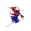
| ||||||||
| Unit cell |
| ||||||||
| Components on special symmetry positions |
| ||||||||
| Details | The biological assembly is created by applying the operation (-x, y, 1/2 - z) with a shift: 0 0 0 |
-Components
| #1: Protein | Mass: 27575.049 Da / Num. of mol.: 1 / Mutation: delta(228-252), R190A Source method: isolated from a genetically manipulated source Source: (gene. exp.) Thermus aquaticus (bacteria) / Gene: ssb / Plasmid: pETTaqSSB delta 228-252 R190A / Production host: |
|---|---|
| #2: Water | ChemComp-HOH /  Mass: 18.015 Da / Num. of mol.: 167 / Source method: isolated from a natural source / Formula: H2O Mass: 18.015 Da / Num. of mol.: 167 / Source method: isolated from a natural source / Formula: H2O |
-Experimental details
-Experiment
| Experiment | Method: X-RAY DIFFRACTION / Number of used crystals: 1 |
|---|
- Sample preparation
| Crystal | Density Matthews: 2.41 Å3/Da / Density % sol: 48.96 % |
|---|---|
| Crystal grow | Temperature: 293.15 K / pH: 6.2 Details: 100 mM Imidazole, 35% 2-Ethoxyethanol, 220 mM Calcium acetate, pH 6.2, VAPOR DIFFUSION, SITTING DROP, temperature 293.15K, pH 6.20 |
-Data collection
| Diffraction | Mean temperature: 100 K |
|---|---|
| Diffraction source | Source: SYNCHROTRON / Site: MPG/DESY, HAMBURG  / Beamline: BW6 / Wavelength: 1.07 / Beamline: BW6 / Wavelength: 1.07 |
| Detector | Type: ADSC QUANTUM 4 / Detector: CCD / Date: Apr 15, 2005 / Details: MIRRORS |
| Radiation | Monochromator: SI(111) DOUBLE-CRYSTAL MONOCHROMATOR WITH COUPLED ROTATIONAL MOTIONS OF TWO FLAT CRYSTALS Protocol: SINGLE WAVELENGTH / Monochromatic (M) / Laue (L): M / Scattering type: x-ray |
| Radiation wavelength | Wavelength: 1.07 Å / Relative weight: 1 |
| Reflection | Resolution: 1.9→20 Å / Num. obs: 21376 / % possible obs: 99.6 % / Observed criterion σ(I): 3 / Redundancy: 18.4 % / Biso Wilson estimate: 32.6 Å2 / Rmerge(I) obs: 0.044 / Rsym value: 0.103 / Net I/σ(I): 20.8 |
| Reflection shell | Resolution: 1.9→2 Å / Redundancy: 17 % / Rmerge(I) obs: 0.144 / Mean I/σ(I) obs: 8.4 / Rsym value: 0.367 / % possible all: 100 |
- Processing
| Software |
| ||||||||||||||||||||||||||||||||||||||||||||||||||||||||||||
|---|---|---|---|---|---|---|---|---|---|---|---|---|---|---|---|---|---|---|---|---|---|---|---|---|---|---|---|---|---|---|---|---|---|---|---|---|---|---|---|---|---|---|---|---|---|---|---|---|---|---|---|---|---|---|---|---|---|---|---|---|---|
| Refinement | Method to determine structure: MOLECULAR REPLACEMENT Starting model: PDB ENTRY 1SE8 Resolution: 1.9→20 Å / Isotropic thermal model: ISOTROPIC / Cross valid method: THROUGHOUT / σ(F): 0 / Stereochemistry target values: ENGH & HUBER
| ||||||||||||||||||||||||||||||||||||||||||||||||||||||||||||
| Displacement parameters | Biso mean: 35.12 Å2 | ||||||||||||||||||||||||||||||||||||||||||||||||||||||||||||
| Refinement step | Cycle: LAST / Resolution: 1.9→20 Å
| ||||||||||||||||||||||||||||||||||||||||||||||||||||||||||||
| Refine LS restraints |
|
