Resolution: 1.85→1.94 Å / Redundancy: 2.23 % / Rmerge(I) obs: 0.175 / Mean I/σ(I) obs: 3.9 / Num. unique all: 8471 / Rsym value: 0.175 / % possible all: 70.2
-
Processing
Software
Name
Version
Classification
PHASES
phasing
CNS
1.1
refinement
MAR345
345DTB
datacollection
DENZO
datareduction
SCALEPACK
datascaling
Refinement
Method to determine structure: MAD / Resolution: 1.85→8 Å / Rfactor Rfree error: 0.002 / Data cutoff high absF: 208869.75 / Data cutoff low absF: 0 / Isotropic thermal model: RESTRAINED / Cross valid method: THROUGHOUT / σ(F): 2 / σ(I): 1 Details: THIS DEPOSITION UPDATES THE COORDINATES OF 1L8A AFTER VALIDATION THROUGH MOLPROBITY (I. W. DAVIS ET AL., NUCL. ACIDS RES. 32, W615-W619, 2004) AND MINIMIZATION WITH CNS (BRUNGER ET AL. ACTA ...Details: THIS DEPOSITION UPDATES THE COORDINATES OF 1L8A AFTER VALIDATION THROUGH MOLPROBITY (I. W. DAVIS ET AL., NUCL. ACIDS RES. 32, W615-W619, 2004) AND MINIMIZATION WITH CNS (BRUNGER ET AL. ACTA CRYST. D54, 905-921, 1998). THE MAIN DIFFERENCE BETWEEN THIS DEPOSITION AND THE ORIGINAL ONE IS THE INTERCHANGE OF NITROGEN AND OXYGEN ATOMS IN SOME AMIDE SIDE CHAINS, AND INTERCHANGE OF NITROGEN AND CARBON ATOMS IN SOME HISTIDINE SIDE CHAINS. THIS DEPOSITION IS BETTER SUITED FOR DETAILED COMPARISONS WITH THE APO AND INHIBITED STRUCTURES 2G67, 1RP7, 2G25 AND 2G28 AS THOSE SUBSEQUENTLY DETERMINED STRUCTURES WERE ALSO TREATED IN THE SAME MANNER. KEY STRUCTURAL DETAILS AND INTERACTIONS DESCRIBED IN OUR 2002 PAPER IN 'BIOCHEMISTRY' HOWEVER, REMAIN UNCHANGED.
In the structure databanks used in Yorodumi, some data are registered as the other names, "COVID-19 virus" and "2019-nCoV". Here are the details of the virus and the list of structure data.
Jan 31, 2019. EMDB accession codes are about to change! (news from PDBe EMDB page)
EMDB accession codes are about to change! (news from PDBe EMDB page)
The allocation of 4 digits for EMDB accession codes will soon come to an end. Whilst these codes will remain in use, new EMDB accession codes will include an additional digit and will expand incrementally as the available range of codes is exhausted. The current 4-digit format prefixed with “EMD-” (i.e. EMD-XXXX) will advance to a 5-digit format (i.e. EMD-XXXXX), and so on. It is currently estimated that the 4-digit codes will be depleted around Spring 2019, at which point the 5-digit format will come into force.
The EM Navigator/Yorodumi systems omit the EMD- prefix.
Related info.:Q: What is EMD? / ID/Accession-code notation in Yorodumi/EM Navigator
Yorodumi is a browser for structure data from EMDB, PDB, SASBDB, etc.
This page is also the successor to EM Navigator detail page, and also detail information page/front-end page for Omokage search.
The word "yorodu" (or yorozu) is an old Japanese word meaning "ten thousand". "mi" (miru) is to see.
Related info.:EMDB / PDB / SASBDB / Comparison of 3 databanks / Yorodumi Search / Aug 31, 2016. New EM Navigator & Yorodumi / Yorodumi Papers / Jmol/JSmol / Function and homology information / Changes in new EM Navigator and Yorodumi
 Movie
Movie Controller


 Open data
 Basic information
 Components
 Keywords
 Function and homology information
 X-RAY DIFFRACTION /
  Authors
 Citation
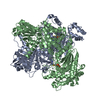
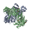
 Structure visualization
 Downloads & links
 Other downloads


 PDBj
PDBj


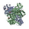
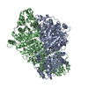
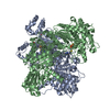
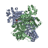
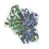
 Assembly
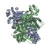

 Mass: 24.305 Da / Num. of mol.: 2 / Source method: obtained synthetically / Formula: Mg
 Mass: 425.314 Da / Num. of mol.: 2 / Source method: obtained synthetically / Formula: C12H19N4O7P2S
 Mass: 18.015 Da / Num. of mol.: 682 / Source method: isolated from a natural source / Formula: H2O
 Sample preparation
 / Beamline: X12C / Wavelength: 1.072
  Processing