 Movie
Movie Controller

[English] 日本語
 Yorodumi
- PDB-2ibu: Crystallographic and kinetic studies of human mitochondrial aceto... -
+ Open data

- Basic information
| Entry | Database: PDB / ID: 2ibu | ||||||
|---|---|---|---|---|---|---|---|
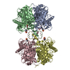
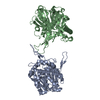
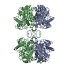
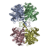
| Title | Crystallographic and kinetic studies of human mitochondrial acetoacetyl-CoA thiolase (T2): the importance of potassium and chloride for its structure and function | ||||||
 Components Components | Acetyl-CoA acetyltransferase | ||||||
 Keywords Keywords | TRANSFERASE / thiolase fold / potassium ion / chloride / beta-alpha-beta-alpha-beta-alpha-beta-beta topology / alpha-beta-alpha-beta-alpha layered structure | ||||||
| Function / homology |  Function and homology information Function and homology informationC-acetyltransferase activity / metanephric proximal convoluted tubule development / propionyl-CoA biosynthetic process / Beta-ketothiolase deficiency / Utilization of Ketone Bodies / Synthesis of Ketone Bodies / cholesterol O-acyltransferase activity / ketone body catabolic process / : / acetyl-CoA catabolic process ...C-acetyltransferase activity / metanephric proximal convoluted tubule development / propionyl-CoA biosynthetic process / Beta-ketothiolase deficiency / Utilization of Ketone Bodies / Synthesis of Ketone Bodies / cholesterol O-acyltransferase activity / ketone body catabolic process / : / acetyl-CoA catabolic process / acetyl-CoA C-acetyltransferase / L-isoleucine catabolic process / coenzyme A binding / Maturation of TCA enzymes and regulation of TCA cycle / acetyl-CoA C-acetyltransferase activity / acetyl-CoA biosynthetic process / Branched-chain amino acid catabolism / coenzyme A metabolic process / coenzyme A biosynthetic process / response to starvation / fatty acid beta-oxidation / potassium ion binding / adipose tissue development / response to hormone / Mitochondrial protein degradation / liver development / mitochondrial matrix / enzyme binding / endoplasmic reticulum / mitochondrion / extracellular exosome / identical protein binding Similarity search - Function | ||||||
| Biological species |  Homo sapiens (human) Homo sapiens (human) | ||||||
| Method |  X-RAY DIFFRACTION / SYNCHROTRON / MOLECULAR REPLACEMENT / Resolution: 1.9 Å X-RAY DIFFRACTION / SYNCHROTRON / MOLECULAR REPLACEMENT / Resolution: 1.9 Å | ||||||
 Authors Authors | Haapalainen, A.M. / Wierenga, R.K. | ||||||
 Citation Citation | Journal: Biochemistry / Year: 2007 Title: Crystallographic and Kinetic Studies of Human Mitochondrial Acetoacetyl-CoA Thiolase: The Importance of Potassium and Chloride Ions for Its Structure and Function Authors: Haapalainen, A.M. / Merilainen, G. / Pirila, P.L. / Kondo, N. / Fukao, T. / Wierenga, R.K. | ||||||
| History |
|
- Structure visualization
| Structure viewer | Molecule: MolmilJmol/JSmol |
|---|
- Downloads & links
-Download
| PDBx/mmCIF format | 2ibu.cif.gz | 325.8 KB | Display | PDBx/mmCIF format |
|---|---|---|---|---|
| PDB format | pdb2ibu.ent.gz | 262.3 KB | Display | PDB format |
| PDBx/mmJSON format | 2ibu.json.gz | Tree view | PDBx/mmJSON format | |
| Others |  Other downloads Other downloads |
-Validation report
| Arichive directory | https://data.pdbj.org/pub/pdb/validation_reports/ib/2ibuftp://data.pdbj.org/pub/pdb/validation_reports/ib/2ibu | HTTPS FTP |
|---|
-Related structure data
| Related structure data |  2ib7C  2ib8C  2ib9C  2ibwC 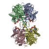 2ibyC  1wl4S C: citing same article ( S: Starting model for refinement |
|---|---|
| Similar structure data |


-Links
 PDBj
PDBj


- Assembly
| Deposited unit | 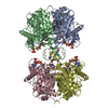
| |||||||||||||||||||||||||||||||||||||||||||||||||||||||||||||||||||||||||||||||||||||||||||||||||||||||||||||||||||||||||||||||||||||||||||||||||||||||||||||||||||||||||||||||||||||||||||||||||||||||||||
|---|---|---|---|---|---|---|---|---|---|---|---|---|---|---|---|---|---|---|---|---|---|---|---|---|---|---|---|---|---|---|---|---|---|---|---|---|---|---|---|---|---|---|---|---|---|---|---|---|---|---|---|---|---|---|---|---|---|---|---|---|---|---|---|---|---|---|---|---|---|---|---|---|---|---|---|---|---|---|---|---|---|---|---|---|---|---|---|---|---|---|---|---|---|---|---|---|---|---|---|---|---|---|---|---|---|---|---|---|---|---|---|---|---|---|---|---|---|---|---|---|---|---|---|---|---|---|---|---|---|---|---|---|---|---|---|---|---|---|---|---|---|---|---|---|---|---|---|---|---|---|---|---|---|---|---|---|---|---|---|---|---|---|---|---|---|---|---|---|---|---|---|---|---|---|---|---|---|---|---|---|---|---|---|---|---|---|---|---|---|---|---|---|---|---|---|---|---|---|---|---|---|---|---|---|
| 1 |
| |||||||||||||||||||||||||||||||||||||||||||||||||||||||||||||||||||||||||||||||||||||||||||||||||||||||||||||||||||||||||||||||||||||||||||||||||||||||||||||||||||||||||||||||||||||||||||||||||||||||||||
| Unit cell |
| |||||||||||||||||||||||||||||||||||||||||||||||||||||||||||||||||||||||||||||||||||||||||||||||||||||||||||||||||||||||||||||||||||||||||||||||||||||||||||||||||||||||||||||||||||||||||||||||||||||||||||
| Noncrystallographic symmetry (NCS) | NCS domain:
NCS domain segments: Component-ID: 1
|