 Movie
Movie Controller

[English] 日本語
 Yorodumi
- PDB-2i3u: Structural studies of protein tyrosine phosphatase beta catalytic... -
+ Open data

- Basic information
| Entry | Database: PDB / ID: 2i3u | ||||||
|---|---|---|---|---|---|---|---|
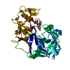
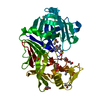
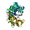
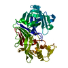
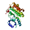
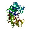
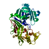
| Title | Structural studies of protein tyrosine phosphatase beta catalytic domain in complex with inhibitors | ||||||
 Components Components | Receptor-type tyrosine-protein phosphatase beta | ||||||
 Keywords Keywords | HYDROLASE / protein tyrosine phosphatase / WPD-loop / sulfamic acid / phosphatase / inhibitor / drug design | ||||||
| Function / homology |  Function and homology information Function and homology informationtransmembrane receptor protein tyrosine kinase inhibitor activity / glial cell migration / transmembrane receptor protein tyrosine phosphatase activity / dephosphorylation / phosphate-containing compound metabolic process / protein dephosphorylation / tertiary granule membrane / specific granule membrane / protein-tyrosine-phosphatase / osteoblast differentiation ...transmembrane receptor protein tyrosine kinase inhibitor activity / glial cell migration / transmembrane receptor protein tyrosine phosphatase activity / dephosphorylation / phosphate-containing compound metabolic process / protein dephosphorylation / tertiary granule membrane / specific granule membrane / protein-tyrosine-phosphatase / osteoblast differentiation / angiogenesis / signaling receptor complex / cadherin binding / Neutrophil degranulation / plasma membrane Similarity search - Function | ||||||
| Biological species |  Homo sapiens (human) Homo sapiens (human) | ||||||
| Method |  X-RAY DIFFRACTION / SYNCHROTRON / MOLECULAR REPLACEMENT / Resolution: 1.85 Å X-RAY DIFFRACTION / SYNCHROTRON / MOLECULAR REPLACEMENT / Resolution: 1.85 Å | ||||||
 Authors Authors | Evdokimov, A.G. / Pokross, M.E. / Walter, R.L. / Mekel, M. | ||||||
 Citation Citation | Journal: Acta Crystallogr.,Sect.D / Year: 2006 Title: Engineering the catalytic domain of human protein tyrosine phosphatase beta for structure-based drug discovery. Authors: Evdokimov, A.G. / Pokross, M. / Walter, R. / Mekel, M. / Cox, B. / Li, C. / Bechard, R. / Genbauffe, F. / Andrews, R. / Diven, C. / Howard, B. / Rastogi, V. / Gray, J. / Maier, M. / Peters, K.G. | ||||||
| History |
|
- Structure visualization
| Structure viewer | Molecule: MolmilJmol/JSmol |
|---|
- Downloads & links
-Download
| PDBx/mmCIF format | 2i3u.cif.gz | 76.8 KB | Display | PDBx/mmCIF format |
|---|---|---|---|---|
| PDB format | pdb2i3u.ent.gz | 56.1 KB | Display | PDB format |
| PDBx/mmJSON format | 2i3u.json.gz | Tree view | PDBx/mmJSON format | |
| Others |  Other downloads Other downloads |
-Validation report
| Arichive directory | https://data.pdbj.org/pub/pdb/validation_reports/i3/2i3uftp://data.pdbj.org/pub/pdb/validation_reports/i3/2i3u | HTTPS FTP |
|---|
-Related structure data
| Related structure data |  2hc1C  2hc2C  2i3rSC  2i4eC 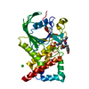 2i4gC 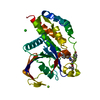 2i4hC 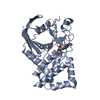 2i5xC C: citing same article ( S: Starting model for refinement |
|---|---|
| Similar structure data |


-Links
 PDBj
PDBj


- Assembly
| Deposited unit | 
| ||||||||
|---|---|---|---|---|---|---|---|---|---|
| 1 |
| ||||||||
| Unit cell |
|
-Components
| #1: Protein | Mass: 36288.035 Da / Num. of mol.: 1 / Fragment: catalytic domain, residues 1662-1973 Source method: isolated from a genetically manipulated source Source: (gene. exp.) Homo sapiens (human) / Gene: PTPRB, PTPB / Plasmid: pDEST-HisMBP / Production host:  |
|---|---|
| #2: Water | ChemComp-HOH /  Mass: 18.015 Da / Num. of mol.: 204 / Source method: isolated from a natural source / Formula: H2O Mass: 18.015 Da / Num. of mol.: 204 / Source method: isolated from a natural source / Formula: H2O |
-Experimental details
-Experiment
| Experiment | Method: X-RAY DIFFRACTION / Number of used crystals: 1 |
|---|
- Sample preparation
| Crystal | Density Matthews: 2.21 Å3/Da / Density % sol: 44.32 % |
|---|---|
| Crystal grow | Temperature: 278 K / Method: vapor diffusion, hanging drop / pH: 8 Details: 21% PEG 8000, 220 mM MgCl2, 1% BME, 0.1% BOG, 5mM DTT, VAPOR DIFFUSION, HANGING DROP, temperature 278K |
-Data collection
| Diffraction | Mean temperature: 100 K |
|---|---|
| Diffraction source | Source: SYNCHROTRON / Site: APS  / Beamline: 22-ID / Wavelength: 1 Å / Beamline: 22-ID / Wavelength: 1 Å |
| Detector | Type: MARRESEARCH / Detector: CCD / Date: Jan 1, 2004 / Details: Si monochromator |
| Radiation | Monochromator: Si / Protocol: SINGLE WAVELENGTH / Monochromatic (M) / Laue (L): M / Scattering type: x-ray |
| Radiation wavelength | Wavelength: 1 Å / Relative weight: 1 |
| Reflection | Resolution: 1.85→22.31 Å / Num. all: 27971 / Num. obs: 27971 / % possible obs: 98.2 % / Observed criterion σ(F): 0 / Observed criterion σ(I): 0 / Redundancy: 4.5 % / Biso Wilson estimate: 26.4 Å2 / Rmerge(I) obs: 0.028 / Net I/σ(I): 21.75 |
| Reflection shell | Resolution: 1.85→1.95 Å / Redundancy: 3.5 % / Rmerge(I) obs: 0.19 / Mean I/σ(I) obs: 5.11 / Num. unique all: 3939 / % possible all: 93.3 |
- Processing
| Software |
| ||||||||||||||||||||||||||||||||||||||||||||||||||||||||||||||||||||||||||||||||||||||||||
|---|---|---|---|---|---|---|---|---|---|---|---|---|---|---|---|---|---|---|---|---|---|---|---|---|---|---|---|---|---|---|---|---|---|---|---|---|---|---|---|---|---|---|---|---|---|---|---|---|---|---|---|---|---|---|---|---|---|---|---|---|---|---|---|---|---|---|---|---|---|---|---|---|---|---|---|---|---|---|---|---|---|---|---|---|---|---|---|---|---|---|---|
| Refinement | Method to determine structure: MOLECULAR REPLACEMENT Starting model: PDB ENTRY 2I3R Resolution: 1.85→22.31 Å / Cor.coef. Fo:Fc: 0.954 / Cor.coef. Fo:Fc free: 0.939 / SU B: 5.312 / SU ML: 0.083 / TLS residual ADP flag: LIKELY RESIDUAL / Isotropic thermal model: TLS / Cross valid method: THROUGHOUT / σ(F): 0 / ESU R: 0.139 / ESU R Free: 0.132 / Stereochemistry target values: MAXIMUM LIKELIHOOD / Details: HYDROGENS HAVE BEEN ADDED IN THE RIDING POSITIONS
| ||||||||||||||||||||||||||||||||||||||||||||||||||||||||||||||||||||||||||||||||||||||||||
| Solvent computation | Ion probe radii: 0.8 Å / Shrinkage radii: 0.8 Å / VDW probe radii: 1.2 Å / Solvent model: MASK | ||||||||||||||||||||||||||||||||||||||||||||||||||||||||||||||||||||||||||||||||||||||||||
| Displacement parameters | Biso mean: 30.832 Å2
| ||||||||||||||||||||||||||||||||||||||||||||||||||||||||||||||||||||||||||||||||||||||||||
| Refinement step | Cycle: LAST / Resolution: 1.85→22.31 Å
| ||||||||||||||||||||||||||||||||||||||||||||||||||||||||||||||||||||||||||||||||||||||||||
| Refine LS restraints |
| ||||||||||||||||||||||||||||||||||||||||||||||||||||||||||||||||||||||||||||||||||||||||||
| LS refinement shell | Resolution: 1.85→1.895 Å / Total num. of bins used: 20
| ||||||||||||||||||||||||||||||||||||||||||||||||||||||||||||||||||||||||||||||||||||||||||
| Refinement TLS params. | Method: refined / Details: Chain A / Origin x: 1.907 Å / Origin y: 21.749 Å / Origin z: 14.919 Å
| ||||||||||||||||||||||||||||||||||||||||||||||||||||||||||||||||||||||||||||||||||||||||||
| Refinement TLS group | Selection: ALL |
