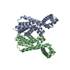
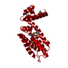
BIOMOLECULE: 1 THIS ENTRY CONTAINS THE CRYSTALLOGRAPHIC ASYMMETRIC UNIT WHICH CONSISTS OF 1 CHAIN. ... BIOMOLECULE: 1 THIS ENTRY CONTAINS THE CRYSTALLOGRAPHIC ASYMMETRIC UNIT WHICH CONSISTS OF 1 CHAIN. SEE REMARK 350 FOR INFORMATION ON GENERATING THE BIOLOGICAL MOLECULE(S). ASSIGNMENT OF A DIMER AS THE BIOLOGICAL UNIT IS SUPPORTED BY SIZE EXCLUSION CHROMATOGRAPHY.
Mass: 18.015 Da / Num. of mol.: 292 / Source method: isolated from a natural source / Formula: H2O
Has protein modification
Y
-
Experimental details
-
Experiment
Experiment
Method: X-RAY DIFFRACTION / Number of used crystals: 1
-
Sample preparation
Crystal
Density Matthews: 2.52 Å3/Da / Density % sol: 51.22 % Description: THE STRUCTURE WAS INITIALLY SOLVED BY MAD FROM ONE CRYSTAL AND REFINEMENT WAS AGAINST DATA FROM A SECOND HIGHER RESOLUTION CRYSTAL
Crystal grow
Temperature: 277 K / Method: vapor diffusion, sitting drop, nanodrop / pH: 6.2 Details: 0.2M NH4F, 20.0% PEG-3350, No Buffer pH 6.2, VAPOR DIFFUSION, SITTING DROP, NANODROP, temperature 277K
Monochromator: Single crystal Si(111) bent monochromator (horizontal focusing) Protocol: MAD / Monochromatic (M) / Laue (L): M / Scattering type: x-ray
Radiation wavelength
ID
Wavelength (Å)
Relative weight
1
1.000001
1
2
0.979197
1
3
0.91837
1
Reflection
Resolution: 1.6→27.126 Å / Num. obs: 27542 / % possible obs: 100 % / Redundancy: 8.3 % / Rmerge(I) obs: 0.083 / Rsym value: 0.083 / Net I/σ(I): 6.1
Reflection shell
Diffraction-ID: 1
Resolution (Å)
Redundancy (%)
Rmerge(I) obs
Mean I/σ(I) obs
Num. measured all
Num. unique all
Rsym value
% possible all
1.64-1.68
5.8
0.798
1
11400
1973
0.798
99.9
1.68-1.73
5.8
0.683
1.1
11503
1989
0.683
99.9
1.73-1.78
5.8
0.515
1.5
11011
1887
0.515
100
1.78-1.83
5.8
0.405
1.9
10865
1862
0.405
100
1.83-1.89
5.9
0.342
2.2
10549
1800
0.342
100
1.89-1.96
5.8
0.253
2.9
10268
1763
0.253
100
1.96-2.03
5.9
0.191
3.9
9768
1669
0.191
100
2.03-2.12
8.4
0.202
3.4
13792
1633
0.202
100
2.12-2.21
11.1
0.173
4
17290
1554
0.173
100
2.21-2.32
11
0.146
4.6
16411
1498
0.146
100
2.32-2.44
11
0.12
5.7
15770
1428
0.12
100
2.44-2.59
11
0.096
6.9
14659
1335
0.096
100
2.59-2.77
10.9
0.085
7.5
13933
1280
0.085
100
2.77-2.99
10.9
0.078
7.6
12719
1172
0.078
100
2.99-3.28
10.8
0.065
9.1
11960
1108
0.065
100
3.28-3.67
10.6
0.06
9.9
10587
996
0.06
100
3.67-4.23
10.1
0.053
11.2
8988
891
0.053
100
4.23-5.19
10.6
0.054
11.1
8090
761
0.054
100
5.19-7.33
10.6
0.051
11.7
6321
599
0.051
100
7.33-28.04
8.6
0.043
12.3
2957
344
0.043
97.8
-
Phasing
Phasing
Method: MAD
-
Processing
Software
Name
Version
Classification
NB
MolProbity
3beta29
modelbuilding
SHELX
phasing
REFMAC
5.2.0019
refinement
SCALA
datascaling
PDB_EXTRACT
2
dataextraction
MOSFLM
datareduction
CCP4
(SCALA)
datascaling
SHELXD
phasing
autoSHARP
phasing
Refinement
Method to determine structure: MAD / Resolution: 1.64→27.126 Å / Cor.coef. Fo:Fc: 0.968 / Cor.coef. Fo:Fc free: 0.96 / SU B: 3.26 / SU ML: 0.057 / TLS residual ADP flag: LIKELY RESIDUAL / Cross valid method: THROUGHOUT / σ(F): 0 / ESU R: 0.085 / ESU R Free: 0.089 Stereochemistry target values: MAXIMUM LIKELIHOOD WITH PHASES Details: 1. HYDROGENS HAVE BEEN ADDED IN THE RIDING POSITIONS. 2. A MET-INHIBITION PROTOCOL WAS USED FOR SELENOMETHIONINE INCORPORATION DURING PROTEIN EXPRESSION. THE OCCUPANCY OF THE SE ATOMS IN THE ...Details: 1. HYDROGENS HAVE BEEN ADDED IN THE RIDING POSITIONS. 2. A MET-INHIBITION PROTOCOL WAS USED FOR SELENOMETHIONINE INCORPORATION DURING PROTEIN EXPRESSION. THE OCCUPANCY OF THE SE ATOMS IN THE MSE RESIDUES WAS REDUCED TO 0.75 FOR THE REDUCED SCATTERING POWER DUE TO PARTIAL S-MET INCORPORATION. 3. EDO MOLECULES FROM THE CRYO SOLUTION ARE MODELED. 4. ATOM RECORDS CONTAIN RESIDUAL B FACTORS ONLY.
Rfactor
Num. reflection
% reflection
Selection details
Rfree
0.201
1386
5 %
RANDOM
Rwork
0.163
-
-
-
obs
0.165
27537
99.93 %
-
Solvent computation
Ion probe radii: 0.8 Å / Shrinkage radii: 0.8 Å / VDW probe radii: 1.2 Å / Solvent model: BABINET MODEL WITH MASK
Displacement parameters
Biso mean: 18.116 Å2
Baniso -1
Baniso -2
Baniso -3
1-
0.61 Å2
0.3 Å2
0 Å2
2-
-
0.61 Å2
0 Å2
3-
-
-
-0.91 Å2
Refinement step
Cycle: LAST / Resolution: 1.64→27.126 Å
Protein
Nucleic acid
Ligand
Solvent
Total
Num. atoms
1462
0
20
292
1774
Refine LS restraints
Refine-ID
Type
Dev ideal
Dev ideal target
Number
X-RAY DIFFRACTION
r_bond_refined_d
0.015
0.022
1577
X-RAY DIFFRACTION
r_bond_other_d
0.002
0.02
1088
X-RAY DIFFRACTION
r_angle_refined_deg
1.355
1.978
2145
X-RAY DIFFRACTION
r_angle_other_deg
0.907
3
2647
X-RAY DIFFRACTION
r_dihedral_angle_1_deg
4.079
5
222
X-RAY DIFFRACTION
r_dihedral_angle_2_deg
33.417
23.649
74
X-RAY DIFFRACTION
r_dihedral_angle_3_deg
11.738
15
276
X-RAY DIFFRACTION
r_dihedral_angle_4_deg
12.119
15
17
X-RAY DIFFRACTION
r_chiral_restr
0.077
0.2
246
X-RAY DIFFRACTION
r_gen_planes_refined
0.006
0.02
1816
X-RAY DIFFRACTION
r_gen_planes_other
0.001
0.02
323
X-RAY DIFFRACTION
r_nbd_refined
0.252
0.2
346
X-RAY DIFFRACTION
r_nbd_other
0.205
0.2
1174
X-RAY DIFFRACTION
r_nbtor_refined
0.181
0.2
784
X-RAY DIFFRACTION
r_nbtor_other
0.089
0.2
831
X-RAY DIFFRACTION
r_xyhbond_nbd_refined
0.168
0.2
202
X-RAY DIFFRACTION
r_symmetry_vdw_refined
0.296
0.2
13
X-RAY DIFFRACTION
r_symmetry_vdw_other
0.24
0.2
61
X-RAY DIFFRACTION
r_symmetry_hbond_refined
0.189
0.2
26
X-RAY DIFFRACTION
r_mcbond_it
2.395
3
1077
X-RAY DIFFRACTION
r_mcbond_other
0.526
3
409
X-RAY DIFFRACTION
r_mcangle_it
2.762
5
1599
X-RAY DIFFRACTION
r_scbond_it
4.658
8
609
X-RAY DIFFRACTION
r_scangle_it
6.475
11
531
LS refinement shell
Resolution: 1.64→1.683 Å / Total num. of bins used: 20
Rfactor
Num. reflection
% reflection
Rfree
0.273
104
-
Rwork
0.216
1880
-
obs
-
1984
99.9 %
Refinement TLS params.
Method: refined / Origin x: 22.46 Å / Origin y: 41.354 Å / Origin z: 12.35 Å
11
12
13
21
22
23
31
32
33
T
-0.0176 Å2
0.0272 Å2
0.0011 Å2
-
0.004 Å2
0.0016 Å2
-
-
0.0344 Å2
L
0.0853 °2
0.1253 °2
-0.1518 °2
-
0.1992 °2
-0.1739 °2
-
-
0.4291 °2
S
0.0353 Å °
-0.0427 Å °
0.0043 Å °
0.0015 Å °
-0.0321 Å °
0.0171 Å °
-0.0022 Å °
0.0392 Å °
-0.0032 Å °
Refinement TLS group
Selection: ALL
+
About Yorodumi
-
News
-
Feb 9, 2022. New format data for meta-information of EMDB entries
New format data for meta-information of EMDB entries
Version 3 of the EMDB header file is now the official format.
The previous official version 1.9 will be removed from the archive.
In the structure databanks used in Yorodumi, some data are registered as the other names, "COVID-19 virus" and "2019-nCoV". Here are the details of the virus and the list of structure data.
Jan 31, 2019. EMDB accession codes are about to change! (news from PDBe EMDB page)
EMDB accession codes are about to change! (news from PDBe EMDB page)
The allocation of 4 digits for EMDB accession codes will soon come to an end. Whilst these codes will remain in use, new EMDB accession codes will include an additional digit and will expand incrementally as the available range of codes is exhausted. The current 4-digit format prefixed with “EMD-” (i.e. EMD-XXXX) will advance to a 5-digit format (i.e. EMD-XXXXX), and so on. It is currently estimated that the 4-digit codes will be depleted around Spring 2019, at which point the 5-digit format will come into force.
The EM Navigator/Yorodumi systems omit the EMD- prefix.
Related info.:Q: What is EMD? / ID/Accession-code notation in Yorodumi/EM Navigator
Yorodumi is a browser for structure data from EMDB, PDB, SASBDB, etc.
This page is also the successor to EM Navigator detail page, and also detail information page/front-end page for Omokage search.
The word "yorodu" (or yorozu) is an old Japanese word meaning "ten thousand". "mi" (miru) is to see.
Related info.:EMDB / PDB / SASBDB / Comparison of 3 databanks / Yorodumi Search / Aug 31, 2016. New EM Navigator & Yorodumi / Yorodumi Papers / Jmol/JSmol / Function and homology information / Changes in new EM Navigator and Yorodumi
 Movie
Movie Controller

 Yorodumi
 Open data
 Basic information
 Components
 Keywords
 Function and homology information
 Pectobacterium atrosepticum (bacteria)
 X-RAY DIFFRACTION /
  Authors
 Citation
 Structure visualization
 Downloads & links
 Other downloads
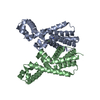


 PDBj
PDBj Assembly
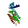

 Mass: 62.068 Da / Num. of mol.: 5 / Source method: obtained synthetically / Formula: C2H6O2
 Mass: 18.015 Da / Num. of mol.: 292 / Source method: isolated from a natural source / Formula: H2O
 Sample preparation
 Processing