 Movie
Movie Controller

[English] 日本語
 Yorodumi
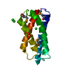
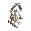
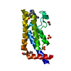
- PDB-2huj: Crystal structure of a protein of uknown function (NP_471338.1) f... -
+ Open data

- Basic information
| Entry | Database: PDB / ID: 2huj | ||||||
|---|---|---|---|---|---|---|---|
| Title | Crystal structure of a protein of uknown function (NP_471338.1) from Listeria innocua at 1.74 A resolution | ||||||
 Components Components | Lin2004 protein | ||||||
 Keywords Keywords | STRUCTURAL GENOMICS / UNKNOWN FUNCTION / NP_471338.1 / hypothetical protein / PSI-2 / Protein Structure Initiative / Joint Center for Structural Genomics / JCSG | ||||||
| Function / homology | YppE-like / YppE-like / YppE-like superfamily / Bacterial domain of unknown function (DUF1798) / Four Helix Bundle (Hemerythrin (Met), subunit A) / Up-down Bundle / Mainly Alpha / Lin2004 protein Function and homology information Function and homology information | ||||||
| Biological species |  Listeria innocua (bacteria) Listeria innocua (bacteria) | ||||||
| Method |  X-RAY DIFFRACTION / SYNCHROTRON / MAD / Resolution: 1.74 Å X-RAY DIFFRACTION / SYNCHROTRON / MAD / Resolution: 1.74 Å | ||||||
 Authors Authors | Joint Center for Structural Genomics (JCSG) | ||||||
 Citation Citation | Journal: Proteins / Year: 2008 Title: Crystal structures of MW1337R and lin2004: representatives of a novel protein family that adopt a four-helical bundle fold. Authors: Kozbial, P. / Xu, Q. / Chiu, H.J. / McMullan, D. / Krishna, S.S. / Miller, M.D. / Abdubek, P. / Acosta, C. / Astakhova, T. / Axelrod, H.L. / Carlton, D. / Clayton, T. / Deller, M. / Duan, L. ...Authors: Kozbial, P. / Xu, Q. / Chiu, H.J. / McMullan, D. / Krishna, S.S. / Miller, M.D. / Abdubek, P. / Acosta, C. / Astakhova, T. / Axelrod, H.L. / Carlton, D. / Clayton, T. / Deller, M. / Duan, L. / Elias, Y. / Elsliger, M.A. / Feuerhelm, J. / Grzechnik, S.K. / Hale, J. / Han, G.W. / Jaroszewski, L. / Jin, K.K. / Klock, H.E. / Knuth, M.W. / Koesema, E. / Kumar, A. / Marciano, D. / Morse, A.T. / Murphy, K.D. / Nigoghossian, E. / Okach, L. / Oommachen, S. / Reyes, R. / Rife, C.L. / Spraggon, G. / Trout, C.V. / van den Bedem, H. / Weekes, D. / White, A. / Wolf, G. / Zubieta, C. / Hodgson, K.O. / Wooley, J. / Deacon, A.M. / Godzik, A. / Lesley, S.A. / Wilson, I.A. | ||||||
| History |
| ||||||
| Remark 999 | SEQUENCE THE CONSTRUCT WAS EXPRESSED WITH A PURIFICATION TAG MGSDKIHHHHHHENLYFQG. |
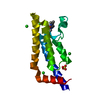
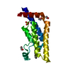
- Structure visualization
| Structure viewer | Molecule: MolmilJmol/JSmol |
|---|
- Downloads & links
-Download
| PDBx/mmCIF format | 2huj.cif.gz | 43.9 KB | Display | PDBx/mmCIF format |
|---|---|---|---|---|
| PDB format | pdb2huj.ent.gz | 30.1 KB | Display | PDB format |
| PDBx/mmJSON format | 2huj.json.gz | Tree view | PDBx/mmJSON format | |
| Others |  Other downloads Other downloads |
-Validation report
| Summary document | 2huj_validation.pdf.gz | 422 KB | Display | wwPDB validaton report |
|---|---|---|---|---|
| Full document | 2huj_full_validation.pdf.gz | 422 KB | Display | |
| Data in XML | 2huj_validation.xml.gz | 7.6 KB | Display | |
| Data in CIF | 2huj_validation.cif.gz | 10.2 KB | Display | |
| Arichive directory | https://data.pdbj.org/pub/pdb/validation_reports/hu/2hujftp://data.pdbj.org/pub/pdb/validation_reports/hu/2huj | HTTPS FTP |
-Related structure data


-Links
 PDBj
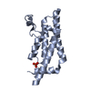
PDBj- Assembly
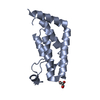
| Deposited unit | 
| ||||||||
|---|---|---|---|---|---|---|---|---|---|
| 1 |
| ||||||||
| Unit cell |
| ||||||||
| Components on special symmetry positions |
|
-Components
| #1: Protein | Mass: 17219.670 Da / Num. of mol.: 1 Source method: isolated from a genetically manipulated source Source: (gene. exp.) Listeria innocua (bacteria) / Gene: NP_471338.1 / Production host: |
|---|---|
| #2: Water | ChemComp-HOH /  Mass: 18.015 Da / Num. of mol.: 97 / Source method: isolated from a natural source / Formula: H2O Mass: 18.015 Da / Num. of mol.: 97 / Source method: isolated from a natural source / Formula: H2O |
| Has protein modification | Y |
-Experimental details
-Experiment
| Experiment | Method: X-RAY DIFFRACTION / Number of used crystals: 1 |
|---|
- Sample preparation
| Crystal |
| |||||||||||||||
|---|---|---|---|---|---|---|---|---|---|---|---|---|---|---|---|---|
| Crystal grow |
|
-Data collection
| Diffraction |
| ||||||||||||||||||||||||||||||||||||||||||||||||||||||||||||||||||||||||||||||||||||||||||||||||||||||||||||||||||||||||||||||||||||||||||||||||||||||||||||||||||||||||
|---|---|---|---|---|---|---|---|---|---|---|---|---|---|---|---|---|---|---|---|---|---|---|---|---|---|---|---|---|---|---|---|---|---|---|---|---|---|---|---|---|---|---|---|---|---|---|---|---|---|---|---|---|---|---|---|---|---|---|---|---|---|---|---|---|---|---|---|---|---|---|---|---|---|---|---|---|---|---|---|---|---|---|---|---|---|---|---|---|---|---|---|---|---|---|---|---|---|---|---|---|---|---|---|---|---|---|---|---|---|---|---|---|---|---|---|---|---|---|---|---|---|---|---|---|---|---|---|---|---|---|---|---|---|---|---|---|---|---|---|---|---|---|---|---|---|---|---|---|---|---|---|---|---|---|---|---|---|---|---|---|---|---|---|---|---|---|---|---|---|
| Diffraction source |
| ||||||||||||||||||||||||||||||||||||||||||||||||||||||||||||||||||||||||||||||||||||||||||||||||||||||||||||||||||||||||||||||||||||||||||||||||||||||||||||||||||||||||
| Detector | Type: ADSC QUANTUM 315 / Detector: CCD / Date: Jul 17, 2006 / Details: Flat mirror (vertical focusing) | ||||||||||||||||||||||||||||||||||||||||||||||||||||||||||||||||||||||||||||||||||||||||||||||||||||||||||||||||||||||||||||||||||||||||||||||||||||||||||||||||||||||||
| Radiation | Monochromator: Single crystal Si(111) bent monochromator (horizontal focusing) Protocol: MAD / Monochromatic (M) / Laue (L): M / Scattering type: x-ray | ||||||||||||||||||||||||||||||||||||||||||||||||||||||||||||||||||||||||||||||||||||||||||||||||||||||||||||||||||||||||||||||||||||||||||||||||||||||||||||||||||||||||
| Radiation wavelength |
| ||||||||||||||||||||||||||||||||||||||||||||||||||||||||||||||||||||||||||||||||||||||||||||||||||||||||||||||||||||||||||||||||||||||||||||||||||||||||||||||||||||||||
| Reflection | Resolution: 1.74→29.424 Å / Num. obs: 20444 / % possible obs: 98.7 % / Redundancy: 11.5 % / Rmerge(I) obs: 0.076 / Rsym value: 0.076 / Net I/σ(I): 6.4 | ||||||||||||||||||||||||||||||||||||||||||||||||||||||||||||||||||||||||||||||||||||||||||||||||||||||||||||||||||||||||||||||||||||||||||||||||||||||||||||||||||||||||
| Reflection shell | Diffraction-ID: 1
|

-Phasing
| Phasing | Method: MAD |
|---|
- Processing
| Software |
| |||||||||||||||||||||||||||||||||||||||||||||||||||||||||||||||||||||||||||||||||||||||||||||||||||||||||||||||||||||||||||||
|---|---|---|---|---|---|---|---|---|---|---|---|---|---|---|---|---|---|---|---|---|---|---|---|---|---|---|---|---|---|---|---|---|---|---|---|---|---|---|---|---|---|---|---|---|---|---|---|---|---|---|---|---|---|---|---|---|---|---|---|---|---|---|---|---|---|---|---|---|---|---|---|---|---|---|---|---|---|---|---|---|---|---|---|---|---|---|---|---|---|---|---|---|---|---|---|---|---|---|---|---|---|---|---|---|---|---|---|---|---|---|---|---|---|---|---|---|---|---|---|---|---|---|---|---|---|---|
| Refinement | Method to determine structure: MAD / Resolution: 1.74→29.424 Å / Cor.coef. Fo:Fc: 0.966 / Cor.coef. Fo:Fc free: 0.953 / SU B: 3.666 / SU ML: 0.059 / TLS residual ADP flag: LIKELY RESIDUAL / Cross valid method: THROUGHOUT / σ(F): 0 / ESU R: 0.086 / ESU R Free: 0.09 Stereochemistry target values: MAXIMUM LIKELIHOOD WITH PHASES Details: (1) HYDROGENS HAVE BEEN ADDED IN THE RIDING POSITIONS. (2) A MET-INHIBITION PROTOCOL WAS USED FOR SELENOMETHIONINE INCORPORATION DURING PROTEIN EXPRESSION. THE OCCUPANCY OF THE SE ATOMS IN ...Details: (1) HYDROGENS HAVE BEEN ADDED IN THE RIDING POSITIONS. (2) A MET-INHIBITION PROTOCOL WAS USED FOR SELENOMETHIONINE INCORPORATION DURING PROTEIN EXPRESSION. THE OCCUPANCY OF THE SE ATOMS IN THE MSE RESIDUES WAS REDUCED TO 0.75 TO ACCOUNT FOR THE REDUCED SCATTERING POWER DUE TO PARTIAL S-MET INCORPORATION.(3) THERE ARE SOME SMALL BLOBS OF UNEXPLAINED ELECTRON DENSITY NEAR RESIDUES A25, A46, A93 AND A96. THEY COULD BE DISORDERED SOLVENT MOLECULES FROM CRYSTALLIZATION SOLUTION. (4) ATOM RECORD CONTAINS RESIDUAL B FACTORS ONLY.
| |||||||||||||||||||||||||||||||||||||||||||||||||||||||||||||||||||||||||||||||||||||||||||||||||||||||||||||||||||||||||||||
| Solvent computation | Ion probe radii: 0.8 Å / Shrinkage radii: 0.8 Å / VDW probe radii: 1.2 Å / Solvent model: BABINET MODEL WITH MASK | |||||||||||||||||||||||||||||||||||||||||||||||||||||||||||||||||||||||||||||||||||||||||||||||||||||||||||||||||||||||||||||
| Displacement parameters | Biso mean: 28.343 Å2
| |||||||||||||||||||||||||||||||||||||||||||||||||||||||||||||||||||||||||||||||||||||||||||||||||||||||||||||||||||||||||||||
| Refinement step | Cycle: LAST / Resolution: 1.74→29.424 Å
| |||||||||||||||||||||||||||||||||||||||||||||||||||||||||||||||||||||||||||||||||||||||||||||||||||||||||||||||||||||||||||||
| Refine LS restraints |
| |||||||||||||||||||||||||||||||||||||||||||||||||||||||||||||||||||||||||||||||||||||||||||||||||||||||||||||||||||||||||||||
| LS refinement shell | Resolution: 1.734→1.779 Å / Total num. of bins used: 20
| |||||||||||||||||||||||||||||||||||||||||||||||||||||||||||||||||||||||||||||||||||||||||||||||||||||||||||||||||||||||||||||
| Refinement TLS params. | Method: refined / Origin x: 28.7236 Å / Origin y: 60.981 Å / Origin z: 11.3069 Å
| |||||||||||||||||||||||||||||||||||||||||||||||||||||||||||||||||||||||||||||||||||||||||||||||||||||||||||||||||||||||||||||
| Refinement TLS group | Selection: ALL |
