 Movie
Movie Controller

[English] 日本語
 Yorodumi
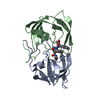
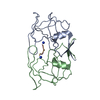
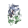
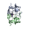
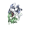
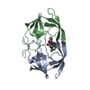
- PDB-2hpe: COMPARISON OF THE STRUCTURES OF HIV-2 PROTEASE COMPLEXES IN THREE... -
+ Open data

- Basic information
| Entry | Database: PDB / ID: 2hpe | ||||||
|---|---|---|---|---|---|---|---|
| Title | COMPARISON OF THE STRUCTURES OF HIV-2 PROTEASE COMPLEXES IN THREE CRYSTAL SPACE GROUPS WITH AN HIV-1 PROTEASE COMPLEX STRUCTURE | ||||||
 Components Components |
| ||||||
 Keywords Keywords | HYDROLASE(ACID PROTEASE) | ||||||
| Function / homology |  Function and homology information Function and homology informationHIV-2 retropepsin / retroviral ribonuclease H / exoribonuclease H / exoribonuclease H activity / host multivesicular body / DNA integration / viral genome integration into host DNA / RNA-directed DNA polymerase / establishment of integrated proviral latency / viral penetration into host nucleus ...HIV-2 retropepsin / retroviral ribonuclease H / exoribonuclease H / exoribonuclease H activity / host multivesicular body / DNA integration / viral genome integration into host DNA / RNA-directed DNA polymerase / establishment of integrated proviral latency / viral penetration into host nucleus / RNA stem-loop binding / RNA-directed DNA polymerase activity / RNA-DNA hybrid ribonuclease activity / Transferases; Transferring phosphorus-containing groups; Nucleotidyltransferases / host cell / viral nucleocapsid / DNA recombination / DNA-directed DNA polymerase / aspartic-type endopeptidase activity / Hydrolases; Acting on ester bonds / DNA-directed DNA polymerase activity / symbiont-mediated suppression of host gene expression / viral translational frameshifting / lipid binding / symbiont entry into host cell / host cell nucleus / host cell plasma membrane / virion membrane / structural molecule activity / proteolysis / DNA binding / zinc ion binding / membrane Similarity search - Function | ||||||
| Biological species |  Human immunodeficiency virus 2 Human immunodeficiency virus 2 | ||||||
| Method |  X-RAY DIFFRACTION / Resolution: 2 Å X-RAY DIFFRACTION / Resolution: 2 Å | ||||||
 Authors Authors | Mulichak, A.M. / Watenpaugh, K.D. | ||||||
 Citation Citation | Journal: To be Published Title: Comparison of the Structures of HIV-2 Protease Complexes in Three Crystal Space Groups with an HIV-1 Protease Complex Structure Authors: Mulichak, A.M. / Watenpaugh, K.D. #1: Journal: J.Biol.Chem. / Year: 1993Title: The Crystallographic Structure of the Protease from Human Immunodeficiency Virus Type 2 with Two Synthetic Peptidic Transition State Analog Inhibitors Authors: Mulichak, A.M. / Hui, J.O. / Tomasselli, A.G. / Heinrikson, R.L. / Curry, K.A. / Tomich, C.-S. / Thaisrivongs, S. / Sawyer, T.K. / Watenpaugh, K.D. | ||||||
| History |
|
- Structure visualization
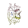
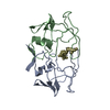
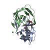
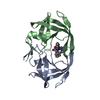
| Structure viewer | Molecule: MolmilJmol/JSmol |
|---|
- Downloads & links
-Download
| PDBx/mmCIF format | 2hpe.cif.gz | 52.1 KB | Display | PDBx/mmCIF format |
|---|---|---|---|---|
| PDB format | pdb2hpe.ent.gz | 37.8 KB | Display | PDB format |
| PDBx/mmJSON format | 2hpe.json.gz | Tree view | PDBx/mmJSON format | |
| Others |  Other downloads Other downloads |
-Validation report
| Summary document | 2hpe_validation.pdf.gz | 384.4 KB | Display | wwPDB validaton report |
|---|---|---|---|---|
| Full document | 2hpe_full_validation.pdf.gz | 391.1 KB | Display | |
| Data in XML | 2hpe_validation.xml.gz | 6.6 KB | Display | |
| Data in CIF | 2hpe_validation.cif.gz | 10.4 KB | Display | |
| Arichive directory | https://data.pdbj.org/pub/pdb/validation_reports/hp/2hpeftp://data.pdbj.org/pub/pdb/validation_reports/hp/2hpe | HTTPS FTP |
-Related structure data


-Links
 PDBj
PDBj
- Assembly
| Deposited unit | 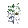
| ||||||||
|---|---|---|---|---|---|---|---|---|---|
| 1 |
| ||||||||
| Unit cell |
|
-Components
| #1: Protein | Mass: 10712.315 Da / Num. of mol.: 2 Source method: isolated from a genetically manipulated source Source: (gene. exp.) Human immunodeficiency virus 2 / Genus: Lentivirus / Production host:  #2: Protein/peptide | | Mass: 783.958 Da / Num. of mol.: 1 Source method: isolated from a genetically manipulated source #3: Water | ChemComp-HOH / |  Mass: 18.015 Da / Num. of mol.: 160 / Source method: isolated from a natural source / Formula: H2O Mass: 18.015 Da / Num. of mol.: 160 / Source method: isolated from a natural source / Formula: H2OSequence details | THERE IS A PEPTIDE FRAGMENT IN THE SUBSTRATE BINDING POCKET. BECAUSE THE SIDE GROUPS ARE NOT KNOWN ...THERE IS A PEPTIDE FRAGMENT IN THE SUBSTRATE BINDING POCKET. BECAUSE THE SIDE GROUPS ARE NOT KNOWN FOR CERTAIN, THE RESIDUES HAVE ALL BEEN REPRESENTE | |
|---|
-Experimental details
-Experiment
| Experiment | Method: X-RAY DIFFRACTION |
|---|
- Sample preparation
| Crystal | Density Matthews: 2.16 Å3/Da / Density % sol: 43.08 % |
|---|
-Data collection
| Reflection | Resolution: 1→10 Å / Num. obs: 10638 / % possible obs: 85 % / Observed criterion σ(F): 2 |
|---|
- Processing
| Software | Name: PROLSQ / Classification: refinement | ||||||||||||
|---|---|---|---|---|---|---|---|---|---|---|---|---|---|
| Refinement | Resolution: 2→10 Å / σ(F): 2 Details: ATOMS WITH B-FACTORS GREATER THAN 70.0 A**2 MAY BE CONSIDERED TO BE DISORDERED OR NOT SEEN IN THE ELECTRON DENSITY MAPS.
| ||||||||||||
| Refinement step | Cycle: LAST / Resolution: 2→10 Å
|
